 |
|
| 包装: |
10 mg |
| 运保温度: |
2-8°C |
| 到货周期: |
登录后查看 |
| 标准价: |
¥客户可见 |
| 会员价: |
¥客户可见 |
| 积分: |
客户可见 分 |
| |
|
运费与支付说明:
1.含干冰类产品有运费;
2. 必须现金付款或有信用额度的会员才可以直接发货,否则需要等待现金付款信息。 |
|
描述:
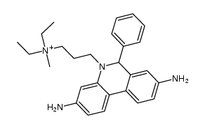
2,7-Diamino-9-phenyl-10 (diethylaminopropyl)-phenanthridium iodide methiodide
Red powder
Reagent for the fluorescent staining of nucleic acids.
POSSIBLE CARCINOGEN!
Description: Reagent used for the fluorescent staining of nucleic acids. Propidium iodide (PI) binds to DNA by intercalating between the bases with little or no sequence preference and with a stoichiometry of one dye per 4-5 base pairs of DNA.1 PI also binds to RNA, necessitating treatment with nucleases to distinguish between RNA and DNA staining. Once the dye is bound to nucleic acids, its fluorescence is enhanced 20- to 30-fold, the fluorescence excitation maximum is shifted ~30-40 nm to the red and the fluorescence emission maximum is shifted ~15 nm to the blue.2 Although its molar absorptivity (extinction coefficient) is relatively low, PI exhibits a sufficiently large Stokes shift to allow simultaneous detection of nuclear DNA and fluorescein-labeled antibodies, provided the proper optical filters are used. Propidium iodide is suitable for fluorescence microscopy, confocal laser scanning microscopy, flow cytometry and fluorometry.
PI is membrane impermeant and generally excluded from viable cells. PI is commonly used for identifying dead cells in a population and as a counterstain in multicolor fluorescent techniques. The counterstaining protocols below are compatible with a wide range of cytological labeling techniques -- direct or indirect antibody -- based detection methods, mRNA in situ hybridization or staining with fluorescent reagents specific for cellular structures. These protocols can be modified for tissue staining.
Protocol for Counterstaining Adherent Cells for Fluorescence Microscopy
Sample Preparation
Use the fixation protocol appropriate for your sample. PI staining is normally performed after all other staining. Note that permeabilization of the cells is required for counterstaining with PI.
RNase Treatment
RNase treatment is required if samples are fixed in paraformaldehyde, formaldehyde or glutaraldehyde. If samples are fixed with methanol/acetic acid or acetone, RNase treatment is usually not required.
-
-
Equilibrate the sample briefly in 2X SSC (0.3 M NaCl, 0.03 M sodium citrate, pH 7.0).
-
Incubate in 100 ug/mL DNase-free RNase in 2X SSC for 20 minutes at 37°C.
-
Rinse samples 3 times, 1 minute each, in 2X SSC.
Counterstaining Protocol
-
-
Equilibrate the sample in 2X SSC.
-
Make a 500 nM solution of PI by diluting the 1 mg/mL (1.5 mM) stock solution 1:3000 in 2X SSC. About 300 µL is usually enough stain for one coverslip preparation. Incubate cells, covered with the dilute stain, for 1-5 minutes.
-
Rinse samples several times in 2X SSC. Drain excess buffer from coverslip and mount in a medium with an antifade reagent.
-
View sample using a fluorescence microscope with appropriate filters.
Protocol for Counterstaining Cells in Suspension for Flow Cytometry
Sample Preparation
-
-
Use the fixation protocol appropriate for your sample, or use the following protocol.
-
Collect a volume of cell suspension corresponding to 2 × 10⁵ to 1 × 10⁶ cells. Pellet the cells by centrifugation. Discard the supernatant, tap the tube to resuspend pellet in the residual liquid and add 1 mL of phosphate-buffered saline (PBS) at room temperature.
-
Transfer the full volume of resuspended cells to 4 mL of absolute ethanol at -20°C by pipetting the cell suspension slowly into the ethanol while vortexing at top speed. Leave in ethanol at -20°C for 5 to 15 minutes.
-
Pellet the cells by centrifugation, discard the ethanol, tap the tube to loosen the pellet and add 5 mL PBS at room temperature. Allow cells to rehydrate for 15 minutes.
Counterstaining Protocol
-
-
Make a 3 mM solution of PI by diluting the 1 mg/mL (1.5 mM) stock solution 1:500 in Staining Buffer (100 mM Tris, pH 7.4, 150 mM NaCl, 1 mM CaCl2 , 0.5 mM MgCl2 , 0.1% Nonidet ® P-40). A 1 mL volume will be required for each cell sample.
-
Centrifuge the cell suspension from the last step of the Sample Preparation, discard the supernatant, tap to loosen the pellet and add 1 mL of PI-Staining Buffer. Incubate for 15 minutes at room temperature and analyze by flow cytometry in the presence of the dye. If the cells are to be viewed by fluorescence microscopy, centrifuge the sample, remove the supernatant and resuspend the cells in fresh buffer. Apply a drop of the suspension to a microscope slide, cover with a coverslip and view.
Protocol for Chromosome FISH Counterstaining
Sample Preparation
Prepare the specimen according to standard procedures.3,4 Briefly rinse the final preparations in dH2O before counterstaining to remove residual buffer salts from the slide. Air dry. This final rinse will help reduce nonspecific background staining on the glass.
Counterstaining Protocol
-
-
Make a 1.5 mM PI staining solution by diluting the 1 mg/mL (1.5 mM) stock solution 1:1000 in PBS. Pipet 300 µL of this staining solution directly onto the specimen. If necessary, RNase A (freshly made) may be added to a final concentration of 10 µg/mL. A plastic coverslip can be used to distribute the dye evenly on the slide.
-
Incubate the specimen in the dark for 30 minutes at room temperature, or at 37°C if RNase is included.
-
Remove the coverslip and rinse briefly with PBS or dH2O to remove unbound dye.
-
Remove excess liquid from the slide by gently blotting around the sample with an absorbent tissue. Place a glass coverslip on the slide, and seal the edges with wax or nail polish. Alternatively, the preparation can be mounted in an antifade reagent according to the manufacturer’s directions.
-
View sample using a fluorescence microscope with appropriate filters.
碘化丙啶;PI
用途:一种可对DNA染色的细胞核染色试剂,常用于细胞凋亡检测,是一种溴化乙啶的类似物,在嵌入双链DNA后释放红色荧光。尽管PI不能通过活细胞膜,但却能穿过破损的细胞膜而对核染色。PI经常被用来与Calcein-AM或者FDA等荧光探针一起使用,能同时对活细胞和死细胞染色。
原厂资料:
2,7-Diamino-9-phenyl-10 (diethylaminopropyl)-phenanthridium iodide methiodide
Red powder
Reagent for the fluorescent staining of nucleic acids.
POSSIBLE CARCINOGEN!
Description: Reagent used for the fluorescent staining of nucleic acids. Propidium iodide (PI) binds to DNA by intercalating between the bases with little or no sequence preference and with a stoichiometry of one dye per 4-5 base pairs of DNA.1 PI also binds to RNA, necessitating treatment with nucleases to distinguish between RNA and DNA staining. Once the dye is bound to nucleic acids, its fluorescence is enhanced 20- to 30-fold, the fluorescence excitation maximum is shifted ~30-40 nm to the red and the fluorescence emission maximum is shifted ~15 nm to the blue.2 Although its molar absorptivity (extinction coefficient) is relatively low, PI exhibits a sufficiently large Stokes shift to allow simultaneous detection of nuclear DNA and fluorescein-labeled antibodies, provided the proper optical filters are used. Propidium iodide is suitable for fluorescence microscopy, confocal laser scanning microscopy, flow cytometry and fluorometry.
PI is membrane impermeant and generally excluded from viable cells. PI is commonly used for identifying dead cells in a population and as a counterstain in multicolor fluorescent techniques. The counterstaining protocols below are compatible with a wide range of cytological labeling techniques -- direct or indirect antibody -- based detection methods, mRNA in situ hybridization or staining with fluorescent reagents specific for cellular structures. These protocols can be modified for tissue staining.
Protocol for Counterstaining Adherent Cells for Fluorescence Microscopy
Sample Preparation
Use the fixation protocol appropriate for your sample. PI staining is normally performed after all other staining. Note that permeabilization of the cells is required for counterstaining with PI.
RNase Treatment
RNase treatment is required if samples are fixed in paraformaldehyde, formaldehyde or glutaraldehyde. If samples are fixed with methanol/acetic acid or acetone, RNase treatment is usually not required.
-
-
Equilibrate the sample briefly in 2X SSC (0.3 M NaCl, 0.03 M sodium citrate, pH 7.0).
-
Incubate in 100 ug/mL DNase-free RNase in 2X SSC for 20 minutes at 37°C.
-
Rinse samples 3 times, 1 minute each, in 2X SSC.
Counterstaining Protocol
-
-
Equilibrate the sample in 2X SSC.
-
Make a 500 nM solution of PI by diluting the 1 mg/mL (1.5 mM) stock solution 1:3000 in 2X SSC. About 300 µL is usually enough stain for one coverslip preparation. Incubate cells, covered with the dilute stain, for 1-5 minutes.
-
Rinse samples several times in 2X SSC. Drain excess buffer from coverslip and mount in a medium with an antifade reagent.
-
View sample using a fluorescence microscope with appropriate filters.
Protocol for Counterstaining Cells in Suspension for Flow Cytometry
Sample Preparation
-
-
Use the fixation protocol appropriate for your sample, or use the following protocol.
-
Collect a volume of cell suspension corresponding to 2 × 10⁵ to 1 × 10⁶ cells. Pellet the cells by centrifugation. Discard the supernatant, tap the tube to resuspend pellet in the residual liquid and add 1 mL of phosphate-buffered saline (PBS) at room temperature.
-
Transfer the full volume of resuspended cells to 4 mL of absolute ethanol at -20°C by pipetting the cell suspension slowly into the ethanol while vortexing at top speed. Leave in ethanol at -20°C for 5 to 15 minutes.
-
Pellet the cells by centrifugation, discard the ethanol, tap the tube to loosen the pellet and add 5 mL PBS at room temperature. Allow cells to rehydrate for 15 minutes.
Counterstaining Protocol
-
-
Make a 3 mM solution of PI by diluting the 1 mg/mL (1.5 mM) stock solution 1:500 in Staining Buffer (100 mM Tris, pH 7.4, 150 mM NaCl, 1 mM CaCl2 , 0.5 mM MgCl2 , 0.1% Nonidet ® P-40). A 1 mL volume will be required for each cell sample.
-
Centrifuge the cell suspension from the last step of the Sample Preparation, discard the supernatant, tap to loosen the pellet and add 1 mL of PI-Staining Buffer. Incubate for 15 minutes at room temperature and analyze by flow cytometry in the presence of the dye. If the cells are to be viewed by fluorescence microscopy, centrifuge the sample, remove the supernatant and resuspend the cells in fresh buffer. Apply a drop of the suspension to a microscope slide, cover with a coverslip and view.
Protocol for Chromosome FISH Counterstaining
Sample Preparation
Prepare the specimen according to standard procedures.3,4 Briefly rinse the final preparations in dH2O before counterstaining to remove residual buffer salts from the slide. Air dry. This final rinse will help reduce nonspecific background staining on the glass.
Counterstaining Protocol
-
-
Make a 1.5 mM PI staining solution by diluting the 1 mg/mL (1.5 mM) stock solution 1:1000 in PBS. Pipet 300 µL of this staining solution directly onto the specimen. If necessary, RNase A (freshly made) may be added to a final concentration of 10 µg/mL. A plastic coverslip can be used to distribute the dye evenly on the slide.
-
Incubate the specimen in the dark for 30 minutes at room temperature, or at 37°C if RNase is included.
-
Remove the coverslip and rinse briefly with PBS or dH2O to remove unbound dye.
-
Remove excess liquid from the slide by gently blotting around the sample with an absorbent tissue. Place a glass coverslip on the slide, and seal the edges with wax or nail polish. Alternatively, the preparation can be mounted in an antifade reagent according to the manufacturer’s directions.
-
View sample using a fluorescence microscope with appropriate filters.
碘化丙啶;PI
用途:一种可对DNA染色的细胞核染色试剂,常用于细胞凋亡检测,是一种溴化乙啶的类似物,在嵌入双链DNA后释放红色荧光。尽管PI不能通过活细胞膜,但却能穿过破损的细胞膜而对核染色。PI经常被用来与Calcein-AM或者FDA等荧光探针一起使用,能同时对活细胞和死细胞染色。
注意事项:
Caution: PI is a known mutagen. Solutions containing PI should be poured through activated charcoal before disposal. The charcoal must then be incinerated to destroy the dye.
Recommended Storage: Upon receipt, store the solid at +4oC, protected from light. The solid should be stable for at least a year. Store the solution of PI at 4°C, protected from light.
Fluorescence Spectral Characteristics: When bound to nucleic acids, the excitation maximum for PI is 535 nm and the emission maximum is 617 nm. PI can be excited with a xenon or mercury-arc lamp or with the 488 line of an argon-ion laser. Generally, PI fluorescence is detected in the FL2 channel of flow cytometers.


 京公网安备11010802025653 版权所有:北京逸优科技有限公司
