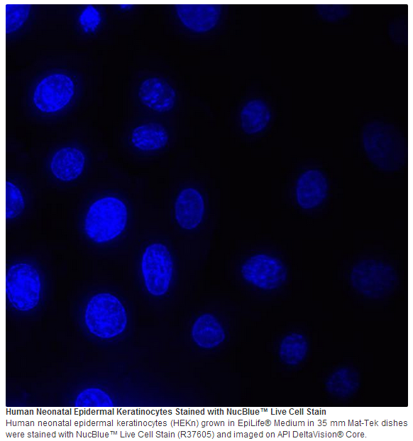
Hoechst 33342 is a popular cell-permeant nuclear counterstain that emits blue fluorescence when bound to DNA. With NucBlue® Live ReadyProbes® Reagent we have formulated this classic stain in a room temperature-stable solution that is provided in a convenient-to-use dropper bottle. Just tip and drip two drops per ml to stain your cells.
• No need to dilute, weigh, or pipette • Convenient dropper bottle—just use two drops per mL • Stable at room temperature—keep handy at your scope or cell culture area • Excited by UV light and emits blue fluorescence at 460 nm when bound to DNA nm
Cell imaging applications The spectral properties of Hoechst 33342 (2'-[4-ethoxyphenyl]-5-[4-methyl-1-piperazinyl]-2,5'-bi-1H-benzimidazole), including a large Stokes shift, make it ideal for use with green (Alexa Fluor® 488, FITC, GFP) and red (Alexa Fluor® 594, Texas Red®, rhodamine, mCherry, mKate-2) fluorophores in multicolor experiments. Because of its high affinity to DNA, Hoechst 33342 is also frequently used in cell counting, cell cycle, and cell replication studies to distinguish condensed nuclei in apoptotic cells, for cell-cycle studies in combination with Click-iT® EdU or BrdU staining, as a nuclear segmentation tool in high content imaging analysis, and to sort cells based on their DNA content..
Suggestions for use NucBlue® Live Cell Stain may be added directly to cells in full media, or buffer solutions. • In most cases 2 drops/ml and an incubation of 15 to 30 minutes will give bright nuclear staining; however, optimization may be needed for some cell types, conditions, and applications. In such cases simply add more, or fewer, drops until the optimal staining intensity is obtained. In most cases, staining intensity increases with time if cells are not washed prior to imaging. • NucBlue® Live Cell Stain is excited by UV light at 360 nm when bound to DNA, with an emission maximum at 460 nm. It is detected through a blue/cyan filter, such as a DAPI filter, blue GFP filters, or the Semrock BrightLine® Alexa Fluor® 350 Dye filter set.
原厂资料:
注意事项:
For Research Use Only. Not for use in diagnostic procedures.


 京公网安备11010802025653 版权所有:北京逸优科技有限公司
