apoptosis-associated speck-like protein containing a CARD
caspase recruitment domain protein 5
Background Information
Apoptosis is regulated by death domain (DD) and/or caspase recruitment domain (CARD) containing molecules and a caspase family of proteases. CARD containing cell death regulators include RAIDD, RICK, Bcl10, Apaf-1, ARC, caspase-2 and caspase-9. A novel CARD domain containing protein has been identified in human and mouse and designated ASC and TMS1. Irregular expression of ASC/TMS1 induced apoptosis through activation of caspase-9 and inhibited the survival of human breast cancer cells. Overexpression of ASC/TMS1 induced DNA fragmentation. ASC/TMS1 is expressed in a variety of human and mouse tissues.
Product Information
Format
Purified
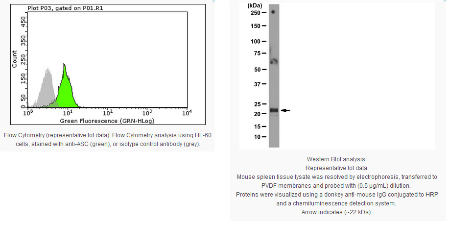
Control
Mouse spleen tissue lysate
Presentation
Purified mouse monoclonal IgG1κ in buffer containing 0.1 M Tris-Glycine (pH 7.4, 150 mM NaCl) with 0.05% sodium azide.
Applications
Application
Detect ASC using this Anti-ASC Antibody, clone 2EI-7 validated for use in WB, FC.
Key Applications
Flow Cytometry
Western Blotting
Biological Information
Immunogen
Full length recombinant human ASC.
Epitope
Unknown
Clone
2EI-7
Concentration
Please refer to the Certificate of Analysis for the lot-specific concentration.
Host
Mouse
Specificity
This antibody recognizes ASC.
Isotype
IgG1κ
Species Reactivity
Human Mouse
Species Reactivity Note
Tested on mouse.
Predicted to react with human based on 100% sequence homology.
This gene encodes an adaptor protein that is composed of two protein-protein interaction domains: a N-terminal PYRIN-PAAD-DAPIN domain (PYD) and a C-terminal caspase-recruitment domain (CARD). The PYD and CARD domains are members of the six-helix bundle death domain-fold superfamily that mediates assembly of large signaling complexes in the inflammatory and apoptotic signaling pathways via the activation of caspase. In normal cells, this protein is localized to the cytoplasm; however, in cells undergoing apoptosis, it forms ball-like aggregates near the nuclear periphery. Two transcript variants encoding different isoforms have been found for this gene.
FUNCTION: Promotes caspase-mediated apoptosis. This proapoptotic activity is mediated predominantly through the activation of caspase 9. May be a component of the inflammasome, a protein complex which also includes NALP2, CARD8 and CASP1 and whose function would be the activation of proinflammatory caspases.
SIZE: 195 amino acids; 21627 Da
SUBUNIT: Forms complexes with other DAPIN domain-containing proteins. Interacts with CIAS1/PYPAF1 and PYDC1.
SUBCELLULAR LOCATION: Cytoplasm. Note=Upstream of caspase activation, a redistribution from the cytoplasm to the aggregates occurs. These appear as hollow, perinuclear spherical, ball-like structures.
TISSUE SPECIFICITY: Widely expressed at low levels. Detected in peripheral blood leukocytes, lung, small intestine, spleen, thymus, colon and at lower levels in placenta, liver and kidney. Very low expression in skeletal muscle, heart and brain. Detected in the leukemia cell lines HL-60 and U937, but not in Jurkat T- cell lymphoma and Daudi Burkitt's lymphoma. Detected in the melanoma cell line WM35, but not in WM793. Not detected in HeLa cervical carcinoma cells and Molt 4 lymphocytic leukemia cells.DOMAIN:SwissProt: Q9ULZ3 Interacts with CIAS1/PYPAF1 and PYDC1 via the DAPIN domain.
MISCELLANEOUS: In breast tumorigenesis, methylation-mediated silencing may affect genes and proteins that act as positive mediators of cell death.
Product Usage Statements
Quality Assurance
Evaluated by Western Blot on mouse spleen tissue lysate.
Western Blot Analysis: 0.5 µg/mL of this antibody detected ASC in mouse spleen tissue lysate.
Usage Statement
Unless otherwise stated in our catalog or other company documentation accompanying the product(s), our products are intended for research use only and are not to be used for any other purpose, which includes but is not limited to, unauthorized commercial uses, in vitro diagnostic uses, ex vivo or in vivo therapeutic uses or any type of consumption or application to humans or animals.
Storage and Shipping Information
Storage Conditions
Stable for 1 year at 2-8°C from date of receipt.
Packaging Information
Material Size
100 µg
原厂资料:
Key Spec Table
Species Reactivity
Key Applications
Host
Format
Antibody Type
H, M
FC, WB
M
Purified
Monoclonal Antibody
Description
Catalogue Number
04-147
Description
Anti-ASC Antibody, clone 2EI-7
Alternate Names
target of methylation-induced silencing-1
Caspase recruitment domain-containing protein 5
PYD and CARD domain containing
PYD and CARD domain-containing protein
Target of methylation-induced silencing 1
apoptosis-associated speck-like protein containing a CARD
caspase recruitment domain protein 5
Background Information
Apoptosis is regulated by death domain (DD) and/or caspase recruitment domain (CARD) containing molecules and a caspase family of proteases. CARD containing cell death regulators include RAIDD, RICK, Bcl10, Apaf-1, ARC, caspase-2 and caspase-9. A novel CARD domain containing protein has been identified in human and mouse and designated ASC and TMS1. Irregular expression of ASC/TMS1 induced apoptosis through activation of caspase-9 and inhibited the survival of human breast cancer cells. Overexpression of ASC/TMS1 induced DNA fragmentation. ASC/TMS1 is expressed in a variety of human and mouse tissues.
Product Information
Format
Purified
Control
Mouse spleen tissue lysate
Presentation
Purified mouse monoclonal IgG1κ in buffer containing 0.1 M Tris-Glycine (pH 7.4, 150 mM NaCl) with 0.05% sodium azide.
Applications
Application
Detect ASC using this Anti-ASC Antibody, clone 2EI-7 validated for use in WB, FC.
Key Applications
Flow Cytometry
Western Blotting
Biological Information
Immunogen
Full length recombinant human ASC.
Epitope
Unknown
Clone
2EI-7
Concentration
Please refer to the Certificate of Analysis for the lot-specific concentration.
Host
Mouse
Specificity
This antibody recognizes ASC.
Isotype
IgG1κ
Species Reactivity
Human Mouse
Species Reactivity Note
Tested on mouse.
Predicted to react with human based on 100% sequence homology.
This gene encodes an adaptor protein that is composed of two protein-protein interaction domains: a N-terminal PYRIN-PAAD-DAPIN domain (PYD) and a C-terminal caspase-recruitment domain (CARD). The PYD and CARD domains are members of the six-helix bundle death domain-fold superfamily that mediates assembly of large signaling complexes in the inflammatory and apoptotic signaling pathways via the activation of caspase. In normal cells, this protein is localized to the cytoplasm; however, in cells undergoing apoptosis, it forms ball-like aggregates near the nuclear periphery. Two transcript variants encoding different isoforms have been found for this gene.
FUNCTION: Promotes caspase-mediated apoptosis. This proapoptotic activity is mediated predominantly through the activation of caspase 9. May be a component of the inflammasome, a protein complex which also includes NALP2, CARD8 and CASP1 and whose function would be the activation of proinflammatory caspases.
SIZE: 195 amino acids; 21627 Da
SUBUNIT: Forms complexes with other DAPIN domain-containing proteins. Interacts with CIAS1/PYPAF1 and PYDC1.
SUBCELLULAR LOCATION: Cytoplasm. Note=Upstream of caspase activation, a redistribution from the cytoplasm to the aggregates occurs. These appear as hollow, perinuclear spherical, ball-like structures.
TISSUE SPECIFICITY: Widely expressed at low levels. Detected in peripheral blood leukocytes, lung, small intestine, spleen, thymus, colon and at lower levels in placenta, liver and kidney. Very low expression in skeletal muscle, heart and brain. Detected in the leukemia cell lines HL-60 and U937, but not in Jurkat T- cell lymphoma and Daudi Burkitt's lymphoma. Detected in the melanoma cell line WM35, but not in WM793. Not detected in HeLa cervical carcinoma cells and Molt 4 lymphocytic leukemia cells.DOMAIN:SwissProt: Q9ULZ3 Interacts with CIAS1/PYPAF1 and PYDC1 via the DAPIN domain.
MISCELLANEOUS: In breast tumorigenesis, methylation-mediated silencing may affect genes and proteins that act as positive mediators of cell death.
Product Usage Statements
Quality Assurance
Evaluated by Western Blot on mouse spleen tissue lysate.
Western Blot Analysis: 0.5 µg/mL of this antibody detected ASC in mouse spleen tissue lysate.
Usage Statement
Unless otherwise stated in our catalog or other company documentation accompanying the product(s), our products are intended for research use only and are not to be used for any other purpose, which includes but is not limited to, unauthorized commercial uses, in vitro diagnostic uses, ex vivo or in vivo therapeutic uses or any type of consumption or application to humans or animals.


 京公网安备11010802025653 版权所有:北京逸优科技有限公司
