细胞成像系统# EVOS? FL Imaging System
- 产品编号:AMF4300-CN 品牌:EVOS-YJ
原厂货号:AMF4300
- 产品分类:
- 应用分类:
 |
|
| 包装: |
台 |
| 运保温度: |
|
| 到货周期: |
登录后查看 |
| 标准价: |
¥客户可见 |
| 会员价: |
¥客户可见 |
| 积分: |
客户可见 分 |
| |
|
运费与支付说明:
1.含干冰类产品有运费;
2. 必须现金付款或有信用额度的会员才可以直接发货,否则需要等待现金付款信息。 |
|
描述:
描述
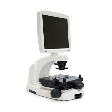
The EVOS® FL Imaging System is a fully integrated, digital, inverted imaging system for four-color fluorescence and transmitted-light applications. It is powerful, yet easy-to-use and delivers high-definition images with exceptional convenience. The unique light cubes, sensitive camera, and precision-engineered optical system make the EVOS® FL system ideal for both demanding and routine fluorescence imaging applications.
The EVOS® FL Imaging System offers you these important advantages:
• Easy installation; no maintenance, assembly, alignment, or calibration
• For four-color fluorescence and transmitted light applications
• Five-position objective turret with front controls
• On-board software
• All-in-one design: digital camera, precision optics, LCD display, and USB storage
Fully Integrated Imaging System
The EVOS® FL system represents a new concept in transmitted light inverted microscopy. It is a fully integrated imaging system that combines precision optics, a 15" high-resolution LCD display, and a highly sensitive Sony ICX445 monochrome CCD camera (1280 x 960 pixel resolution, 1.3 Megapixels). Images are seamlessly acquired through the intuitive user interface using a mouse for easy control. Monochrome cameras are commonly used for high-performance fluorescence applications and provide the best sensitivity for detection of faint fluorescence signals. Color cameras, as on the EVOS® FL Color System, have lower fluorescence sensitivity but have the advantage of being able to differentiate structures by color in transmitted light (e.g., imaging stained tissue samples).
Compare cell imaging systems >
Versatile and Highly Configurable
The EVOS® FL system features an easily accessible 5-position objective turret and a 3-position condenser turret. The 4 fluorescence light cubes are lever-actioned for rapid and easy selection. The optical system can be configured to best meet your needs using our full range of high-quality objectives from 2x to 100x magnification. Lighting settings are automatically adjusted to match the objective magnification. The EVOS® FL system features a mechanical stage with X/Y-axis fine-positioning using control knobs conveniently located on the left side and the front of the stage. The stage accommodates EVOS® vessel holders that provide a perfect fit for most vessel types and sizes. Interchangeable stage plates accommodate most vessel types and sizes, including flasks, Petri dishes, multi-well plates, and slides.
Smart LED Illumination Technology
All EVOS® fluorescence imaging systems utilize our unique, proprietary LED light cubes. This world-leading light engine outputs remarkable intensity over a short light-path that delivers superior fluorophore excitation. Each light cube contains a precisely matched set of optical components to optimize the position, evenness, and intensity of the light beam. Digitally controlled LED light sources allow adjustment of illumination levels, dramatically improving control over photobleaching. Hard-coated filters give sharper edges and significantly higher transmission efficiencies than traditional soft-coated filters.
Integrated Software
The on-board computer makes the EVOS® FL system easy to run and use, and the intuitive, comprehensive software facilitates both image acquisition and analysis. Modules include time-lapse acquisition, manual-assist cell counting, and image review. Saved user settings work in conjunction with "smart" controls to help increase efficiency and ease of use. Image data are saved to a USB flash drive for easy, portable data transfer or to a Windows-networked local server. Images can be saved in PNG, JPEG, BMP, and TIFF formats with a maximum captured image resolution of 1280 x 960 pixels.
These features make the EVOS® FL system ideal for both routine and demanding fluorescence imaging applications such as GFP-vector transfection efficiency analysis, routine cell and tissue culture, cell confluence determination, stem cell growth and differentiation, and developmental biology and tissue slice analyses. And the compact footprint makes it as easy to use the EVOS® FL system in a cell culture hood as on the bench.
Easy to Use and Reliable
The EVOS® FL Imaging System is plug-and-play. In addition to an exceptionally easy set-up and installation, it requires no warm-up or cool-down periods. The LED light source offers exceptional stability and durability—so you can turn the unit on and off whenever you need to image a sample. The environmentally safe mercury-free LED bulbs are rated for >50,000 hours (~17 years) of life, compared to 300 hours for a typical mercury bulb and 1,500 hours for a metal halide bulb. The long life and low energy consumption translate into significantly lower operating costs compared to instruments with conventional light sources. Finally, the advanced ergonomic design of the EVOS® systems removes the strain associated with conventional microscopes, enables shared viewing, and makes moving the units easy. For extended or precision use, the EVOS® arm rest provides comfortable support.
The EVOS® imaging systems are built from the ground up to maximize performance and optimize workflow. You will be astonished at how easy it is to operate and amazed at how extraordinary your images look on-screen.
Learn more about the EVOS® FL Imaging System
Explore the entire EVOS® line of imaging systems and accessories
System Highlights
Optics: infinity-corrected optical system, RMS-threaded objectives with 45 mm parfocal distance
Illumination: adjustable intensity LED (>50,000-hour life per light cube)
Light cubes (not included): broad selection of patented standard and specialty light cubes
Common light cubes include: DAPI (Ex 360 nm/Em 447 nm), GFP (Ex 470 nm/Em 525 nm), RFP (Ex 530 nm/Em 593 nm), Texas Red® (Ex 585 nm/Em 624), Cy5 (Ex 628 nm/Em 692 nm)
Contrast methods: fluorescence and transmitted light (brightfield & phase contrast)
Objective turret: 5-position, front-mounted control
Objectives (not included): wide selection of high-quality LWD and coverslip-corrected objectives available
Condenser: 3-position turret with brightfield and phase contrast annuli
Condenser working distance: 60 mm
Stage: mechanical "glide" stage; X-Y axis fine-positioning controls: 28.3 mm (1.11") per rotation, 110 mm x 110 mm (4.3" x 4.3") range of motion; Z-axis focusing controls: 480 µm/rotation; interchangeable vessel holders are available for most common shapes and sizes
Focus mechanism: coaxial focus knobs with tension control
Course focus: 38 mm/rev
Fine focus: 0.2mm/rev, precision 0.002 mm
LCD display: 15-inch color, 1024 x 768 pixels, adjustable tilt
Camera: Sony ICX445 monochrome CCD, 1/3" 1280 x 960 pixels, 1.3 Megapixels
Image acquisition: onboard microprocessor, built-in software for image acquisition via mouse control
Captured images: 16-bit monochrome TIFF or PNG (12-bit dynamic range); 24-bit color TIFF or PNG; jpeg, bmp, 1280 x 960 pixels
Output ports: 3 USB and 1 DVI
Power supply: AC adapter with country-specific power cords; input 100-240 V, 50-60 Hz; output 12 VDC/4.15 A
Dimensions: operating height = 57.8 cm (22.75 in); storage/transport height = 32.4 cm (12.75 in); depth = 47.0 cm (18.5 in); width = 35.5 cm (14.0 in)
Weight: 15.3 kg (33.7 lbs)
原厂资料:
描述
The EVOS® FL Imaging System is a fully integrated, digital, inverted imaging system for four-color fluorescence and transmitted-light applications. It is powerful, yet easy-to-use and delivers high-definition images with exceptional convenience. The unique light cubes, sensitive camera, and precision-engineered optical system make the EVOS® FL system ideal for both demanding and routine fluorescence imaging applications.
The EVOS® FL Imaging System offers you these important advantages:
• Easy installation; no maintenance, assembly, alignment, or calibration
• For four-color fluorescence and transmitted light applications
• Five-position objective turret with front controls
• On-board software
• All-in-one design: digital camera, precision optics, LCD display, and USB storage
Fully Integrated Imaging System
The EVOS® FL system represents a new concept in transmitted light inverted microscopy. It is a fully integrated imaging system that combines precision optics, a 15" high-resolution LCD display, and a highly sensitive Sony ICX445 monochrome CCD camera (1280 x 960 pixel resolution, 1.3 Megapixels). Images are seamlessly acquired through the intuitive user interface using a mouse for easy control. Monochrome cameras are commonly used for high-performance fluorescence applications and provide the best sensitivity for detection of faint fluorescence signals. Color cameras, as on the EVOS® FL Color System, have lower fluorescence sensitivity but have the advantage of being able to differentiate structures by color in transmitted light (e.g., imaging stained tissue samples).
Compare cell imaging systems >
Versatile and Highly Configurable
The EVOS® FL system features an easily accessible 5-position objective turret and a 3-position condenser turret. The 4 fluorescence light cubes are lever-actioned for rapid and easy selection. The optical system can be configured to best meet your needs using our full range of high-quality objectives from 2x to 100x magnification. Lighting settings are automatically adjusted to match the objective magnification. The EVOS® FL system features a mechanical stage with X/Y-axis fine-positioning using control knobs conveniently located on the left side and the front of the stage. The stage accommodates EVOS® vessel holders that provide a perfect fit for most vessel types and sizes. Interchangeable stage plates accommodate most vessel types and sizes, including flasks, Petri dishes, multi-well plates, and slides.
Smart LED Illumination Technology
All EVOS® fluorescence imaging systems utilize our unique, proprietary LED light cubes. This world-leading light engine outputs remarkable intensity over a short light-path that delivers superior fluorophore excitation. Each light cube contains a precisely matched set of optical components to optimize the position, evenness, and intensity of the light beam. Digitally controlled LED light sources allow adjustment of illumination levels, dramatically improving control over photobleaching. Hard-coated filters give sharper edges and significantly higher transmission efficiencies than traditional soft-coated filters.
Integrated Software
The on-board computer makes the EVOS® FL system easy to run and use, and the intuitive, comprehensive software facilitates both image acquisition and analysis. Modules include time-lapse acquisition, manual-assist cell counting, and image review. Saved user settings work in conjunction with "smart" controls to help increase efficiency and ease of use. Image data are saved to a USB flash drive for easy, portable data transfer or to a Windows-networked local server. Images can be saved in PNG, JPEG, BMP, and TIFF formats with a maximum captured image resolution of 1280 x 960 pixels.
These features make the EVOS® FL system ideal for both routine and demanding fluorescence imaging applications such as GFP-vector transfection efficiency analysis, routine cell and tissue culture, cell confluence determination, stem cell growth and differentiation, and developmental biology and tissue slice analyses. And the compact footprint makes it as easy to use the EVOS® FL system in a cell culture hood as on the bench.
Easy to Use and Reliable
The EVOS® FL Imaging System is plug-and-play. In addition to an exceptionally easy set-up and installation, it requires no warm-up or cool-down periods. The LED light source offers exceptional stability and durability—so you can turn the unit on and off whenever you need to image a sample. The environmentally safe mercury-free LED bulbs are rated for >50,000 hours (~17 years) of life, compared to 300 hours for a typical mercury bulb and 1,500 hours for a metal halide bulb. The long life and low energy consumption translate into significantly lower operating costs compared to instruments with conventional light sources. Finally, the advanced ergonomic design of the EVOS® systems removes the strain associated with conventional microscopes, enables shared viewing, and makes moving the units easy. For extended or precision use, the EVOS® arm rest provides comfortable support.
The EVOS® imaging systems are built from the ground up to maximize performance and optimize workflow. You will be astonished at how easy it is to operate and amazed at how extraordinary your images look on-screen.
Learn more about the EVOS® FL Imaging System
Explore the entire EVOS® line of imaging systems and accessories
System Highlights
Optics: infinity-corrected optical system, RMS-threaded objectives with 45 mm parfocal distance
Illumination: adjustable intensity LED (>50,000-hour life per light cube)
Light cubes (not included): broad selection of patented standard and specialty light cubes
Common light cubes include: DAPI (Ex 360 nm/Em 447 nm), GFP (Ex 470 nm/Em 525 nm), RFP (Ex 530 nm/Em 593 nm), Texas Red® (Ex 585 nm/Em 624), Cy5 (Ex 628 nm/Em 692 nm)
Contrast methods: fluorescence and transmitted light (brightfield & phase contrast)
Objective turret: 5-position, front-mounted control
Objectives (not included): wide selection of high-quality LWD and coverslip-corrected objectives available
Condenser: 3-position turret with brightfield and phase contrast annuli
Condenser working distance: 60 mm
Stage: mechanical "glide" stage; X-Y axis fine-positioning controls: 28.3 mm (1.11") per rotation, 110 mm x 110 mm (4.3" x 4.3") range of motion; Z-axis focusing controls: 480 µm/rotation; interchangeable vessel holders are available for most common shapes and sizes
Focus mechanism: coaxial focus knobs with tension control
Course focus: 38 mm/rev
Fine focus: 0.2mm/rev, precision 0.002 mm
LCD display: 15-inch color, 1024 x 768 pixels, adjustable tilt
Camera: Sony ICX445 monochrome CCD, 1/3" 1280 x 960 pixels, 1.3 Megapixels
Image acquisition: onboard microprocessor, built-in software for image acquisition via mouse control
Captured images: 16-bit monochrome TIFF or PNG (12-bit dynamic range); 24-bit color TIFF or PNG; jpeg, bmp, 1280 x 960 pixels
Output ports: 3 USB and 1 DVI
Power supply: AC adapter with country-specific power cords; input 100-240 V, 50-60 Hz; output 12 VDC/4.15 A
Dimensions: operating height = 57.8 cm (22.75 in); storage/transport height = 32.4 cm (12.75 in); depth = 47.0 cm (18.5 in); width = 35.5 cm (14.0 in)
Weight: 15.3 kg (33.7 lbs)


 京公网安备11010802025653 版权所有:北京逸优科技有限公司
