The LIVE/DEAD® Cell Imaging Kit is a sensitive two-color fluorescence cell viability assay optimized for FITC and Texas Red® filters. Quick and easy to use, the kit allows discrimination between live and dead cells with two probes that measure recognized parameters of cytotoxicity and cell viability—intracellular esterase activity and plasma membrane integrity.
The LIVE/DEAD® Cell Imaging Kit *488/570* offers:
Fast, simple determination of live and dead cells
• Accuracy with convenience
• Sensitive probes ideal for FITC and Texas Red filters
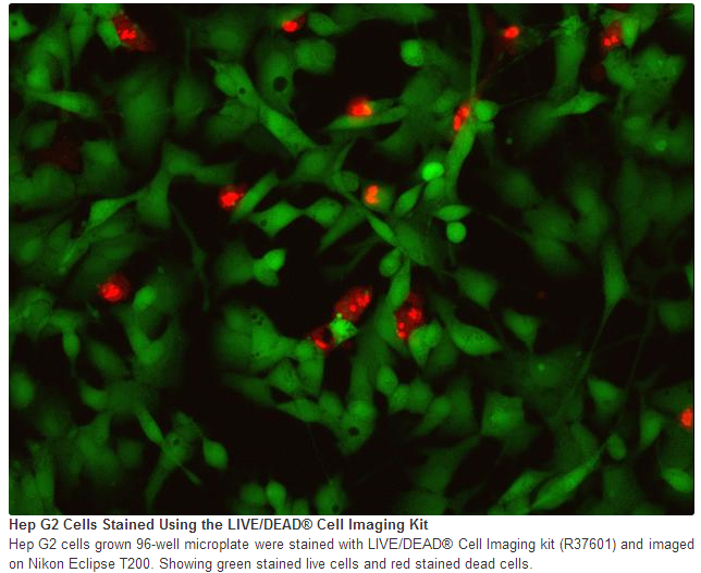
Dual Probe-Based Assay for Imaging Platforms Just like our popular LIVE/DEAD® Viability/Cytotoxicity assay, the new LIVE/DEAD® Cell Imaging Kit is based on a cell-permeable dye for staining of live cells and a cell-impermeable dye for staining of dead and dying cells, which are characterized by compromised cell membranes. To adapt this important assay for imaging platforms, the LIVE/DEAD® Cell Imaging Kit components were optimized for the common green and red imaging filters used with FITC and Texas Red. The live cell component produces an intense, uniform green fluorescence in live cells (ex/em 488 nm/515 nm). The dead cell component produces a predominantly nuclear red fluorescence (ex/em 570nm/602 nm) in cells with compromised cell membranes, a strong indicator of cell death and cytotoxicity (Fig 1).
Quick, Exact Determination of Viability and Cytotoxicity The LIVE/DEAD® Cell Imaging kit provides very sensitive detection of key measures of cell health: viability, live/dead ratio, and cytotoxicity. The components of the LIVE/DEAD® Cell Imaging kit are configured for ease of use with minimal dilutions.
Assay Principle With the LIVE/DEAD® Cell Imaging Kit, live cells are distinguished by the presence of ubiquitous intracellular esterase activity as determined by the enzymatic conversion of the virtually non-fluorescent cell-permeant calcein AM to the intensely fluorescent calcein, which is well-retained within live cells. The red component of the LIVE/DEAD® Cell Imaging Kit is cell-impermeant and therefore only enters cells with damaged membranes. In dying and dead cells a bright red fluorescence is generated upon binding to DNA. Background fluorescence levels are inherently low with this assay technique because the dyes are virtually non-fluorescent before interacting with cells. The fluorophores in the LIVE/DEAD® Cell Imaging Kit were selected for their brightness, spectral properties (FITC and Texas Red filters), and ease of use. Packaged for workflow convenience, they allow for effortless and quick determination of the fractions of live and dead cells in a population.
原厂资料:
注意事项:
For Research Use Only. Not for use in diagnostic procedures.


 京公网安备11010802025653 版权所有:北京逸优科技有限公司
