There are two kinds of IGF receptors based on their relative affinities for IGF1 versus IGF2. The IGF1 receptor prefers IGF1 over IGF2 and weakly binds insulin. IGF1 receptor is a disulfide-linked heterotetrameric transmembrane protein consisting of two alpha (130 kDa) and two beta (95 kDa) subunits. Both the alpha and beta subunits are encoded within a single receptor precursor cDNA. The IGF1 receptor is therefore similar in structure to the insulin receptor. The proreceptor polypeptide is proteolytically cleaved and disulfide-linked to yield the mature heterotetrameric receptor. The IGF1 receptor is highly expressed in all cell types and tissues and is highly overexpressed in most malignant tissues where it functions as an anti-apoptotic agent by enhancing cell survival.
Product Information
Format
Purified
Control
Human placental villi and tonsil tissue
Presentation
Purified mouse monoclonal IgG1κ in buffer containing 0.1 M Tris-Glycine (pH 7.4, 150 mM NaCl) with 0.05% sodium azide.
Applications
Application
This Anti-IGF-1R Antibody, clone 24-57 is validated for use in IH, IP for the detection of IGF-1R.
Key Applications
Immunohistochemistry
Immunoprecipitation
Application Notes
Immunoprecipitation Analysis: A previous lot was used by an independent laboratory in IP. (Soos, M., et al. (1992). Journal of Biological Chemistry. 267(18):12955-12963.)
Biological Information
Immunogen
KLH-conjugated linear peptide corresponding to human IGF-IR.
Epitope
Unknown
Clone
24-57
Concentration
Please refer to the Certificate of Analysis for the lot-specific concentration.
This receptor binds insulin-like growth factor with a high affinity. It has tyrosine kinase activity. The insulin-like growth factor I receptor plays a critical role in transformation events. Cleavage of the precursor generates alpha and beta subunits. It is highly overexpressed in most malignant tissues where it functions as an anti-apoptotic agent by enhancing cell survival. [provided by RefSeq].
FUNCTION: This receptor binds insulin-like growth factor 1 (IGF1) with a high affinity and IGF2 with a lower affinity. It has a tyrosine-protein kinase activity, which is necessary for the activation of the IGF1-stimulated downstream signaling cascade.
SIZE: 1367 amino acids; 154793 Da
SUBUNIT: Tetramer of 2 alpha and 2 beta chains linked by disulfide bonds. The alpha chains contribute to the formation of the ligand- binding domain, while the beta chain carries the kinase domain. Interacts with PIK3R1 and with the PTB/PID domains of IRS1 and SHC1 in vitro when autophosphorylated on tyrosine residues.
SUBCELLULAR LOCATION: Membrane; Single-pass type I membrane protein.
TISSUE SPECIFICITY: Expressed in a variety of tissues.
PTM: The cytoplasmic domain of the beta subunit is autophosphorylated on tyrosine residues in response to insulin- like growth factor I (IGF I). & Phosphorylation of Tyr-980 is required for IRS1- and SHC1- binding.
DISEASE: Defects in IGF1R may be a cause in some cases of resistance to insulin-like growth factor 1 (IGF1 resistance) [MIM:270450]. IGF1 resistance is a growth deficiency disorder characterized by intrauterine growth retardation and poor postnatal growth accompanied with increased plasma IGF1.
SIMILARITY: Belongs to the protein kinase superfamily. Tyr protein kinase family. Insulin receptor subfamily. & Contains 3 fibronectin type-III domains. & Contains 1 protein kinase domain.
Product Usage Statements
Quality Assurance
Evaluated by Immunohistochemistry in human placental villi and tonsil tissue.
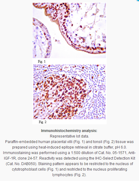
Immunohistochemistry Analysis: A 1:500 dilution from a previous lot detected IGF-1R in human placental villi and tonsil tissue.
Usage Statement
Unless otherwise stated in our catalog or other company documentation accompanying the product(s), our products are intended for research use only and are not to be used for any other purpose, which includes but is not limited to, unauthorized commercial uses, in vitro diagnostic uses, ex vivo or in vivo therapeutic uses or any type of consumption or application to humans or animals.
Storage and Shipping Information
Storage Conditions
Stable for 1 year at 2-8°C from date of receipt.
Packaging Information
Material Size
100 µg
原厂资料:
Key Spec Table
Species Reactivity
Key Applications
Host
Format
Antibody Type
H
IHC, IP
M
Purified
Monoclonal Antibody
Description
Catalogue Number
05-1571
Description
Anti-IGF-1R Antibody, clone 24-57
Alternate Names
CD221 antigen
soluble IGF1R variant 2
soluble IGF1R variant 1
insulin-like growth factor 1 receptor
Insulin-like growth factor I receptor
IGF-I receptor
Background Information
There are two kinds of IGF receptors based on their relative affinities for IGF1 versus IGF2. The IGF1 receptor prefers IGF1 over IGF2 and weakly binds insulin. IGF1 receptor is a disulfide-linked heterotetrameric transmembrane protein consisting of two alpha (130 kDa) and two beta (95 kDa) subunits. Both the alpha and beta subunits are encoded within a single receptor precursor cDNA. The IGF1 receptor is therefore similar in structure to the insulin receptor. The proreceptor polypeptide is proteolytically cleaved and disulfide-linked to yield the mature heterotetrameric receptor. The IGF1 receptor is highly expressed in all cell types and tissues and is highly overexpressed in most malignant tissues where it functions as an anti-apoptotic agent by enhancing cell survival.
Product Information
Format
Purified
Control
Human placental villi and tonsil tissue
Presentation
Purified mouse monoclonal IgG1κ in buffer containing 0.1 M Tris-Glycine (pH 7.4, 150 mM NaCl) with 0.05% sodium azide.
Applications
Application
This Anti-IGF-1R Antibody, clone 24-57 is validated for use in IH, IP for the detection of IGF-1R.
Key Applications
Immunohistochemistry
Immunoprecipitation
Application Notes
Immunoprecipitation Analysis: A previous lot was used by an independent laboratory in IP. (Soos, M., et al. (1992). Journal of Biological Chemistry. 267(18):12955-12963.)
Biological Information
Immunogen
KLH-conjugated linear peptide corresponding to human IGF-IR.
Epitope
Unknown
Clone
24-57
Concentration
Please refer to the Certificate of Analysis for the lot-specific concentration.
This receptor binds insulin-like growth factor with a high affinity. It has tyrosine kinase activity. The insulin-like growth factor I receptor plays a critical role in transformation events. Cleavage of the precursor generates alpha and beta subunits. It is highly overexpressed in most malignant tissues where it functions as an anti-apoptotic agent by enhancing cell survival. [provided by RefSeq].
FUNCTION: This receptor binds insulin-like growth factor 1 (IGF1) with a high affinity and IGF2 with a lower affinity. It has a tyrosine-protein kinase activity, which is necessary for the activation of the IGF1-stimulated downstream signaling cascade.
SIZE: 1367 amino acids; 154793 Da
SUBUNIT: Tetramer of 2 alpha and 2 beta chains linked by disulfide bonds. The alpha chains contribute to the formation of the ligand- binding domain, while the beta chain carries the kinase domain. Interacts with PIK3R1 and with the PTB/PID domains of IRS1 and SHC1 in vitro when autophosphorylated on tyrosine residues.
SUBCELLULAR LOCATION: Membrane; Single-pass type I membrane protein.
TISSUE SPECIFICITY: Expressed in a variety of tissues.
PTM: The cytoplasmic domain of the beta subunit is autophosphorylated on tyrosine residues in response to insulin- like growth factor I (IGF I). & Phosphorylation of Tyr-980 is required for IRS1- and SHC1- binding.
DISEASE: Defects in IGF1R may be a cause in some cases of resistance to insulin-like growth factor 1 (IGF1 resistance) [MIM:270450]. IGF1 resistance is a growth deficiency disorder characterized by intrauterine growth retardation and poor postnatal growth accompanied with increased plasma IGF1.
SIMILARITY: Belongs to the protein kinase superfamily. Tyr protein kinase family. Insulin receptor subfamily. & Contains 3 fibronectin type-III domains. & Contains 1 protein kinase domain.
Product Usage Statements
Quality Assurance
Evaluated by Immunohistochemistry in human placental villi and tonsil tissue.
Immunohistochemistry Analysis: A 1:500 dilution from a previous lot detected IGF-1R in human placental villi and tonsil tissue.
Usage Statement
Unless otherwise stated in our catalog or other company documentation accompanying the product(s), our products are intended for research use only and are not to be used for any other purpose, which includes but is not limited to, unauthorized commercial uses, in vitro diagnostic uses, ex vivo or in vivo therapeutic uses or any type of consumption or application to humans or animals.


 京公网安备11010802025653 版权所有:北京逸优科技有限公司
