PDGFR (Platelet-Derived Growth Factor Receptor) is a receptor tyrosine kinase for the PDGF family of growth factors. PDGF homo and hetero- dimmers (dimer of A (17 kDa) and B (14 kDa) chains (AA, AB, or BB)) bind to the two structurally related a and b PDGF Receptors (PDGFRa and PDGFRb). The type A PDGF Receptor binds all three PDGFs with high affinity, whereas the type B PDGF Receptor is selective for PDGF-BB. This binding results in the dimerization of the receptor and the subsequent kinase activation of the receptor tyrosine kinase (RTK) and its autophosphorylation. These phosphorylations allow for various proteins, such as PI3 Kinase, PLCg, and Grb2, to bind through their SH2 domains and transduce signals into the cell. The activation of PDGFFR triggers the intracellular loop and activates Ras, Erk1/2, and ROS. PDGFb has been implicated in embryonic development, angiogenesis, and development of tumors of glial origin.
Product Information
Format
Purified
Control
Jurkat cell lysate
Presentation
Purified mouse monoclonal IgG1 in buffer containing 20 mM sodium phosphate, 250 mM NaCl, 0.1% NaN3, pH. 7.6
Applications
Application
This Anti-PDGFR Antibody, clone CH-3 is validated for use in WB, ELISA, IH(P) for the detection of PDGFR.
Key Applications
Western Blotting
Immunohistochemistry (Paraffin)
ELISA
Application Notes
ELISA: A previous lot of this antibody was used in ELISA.
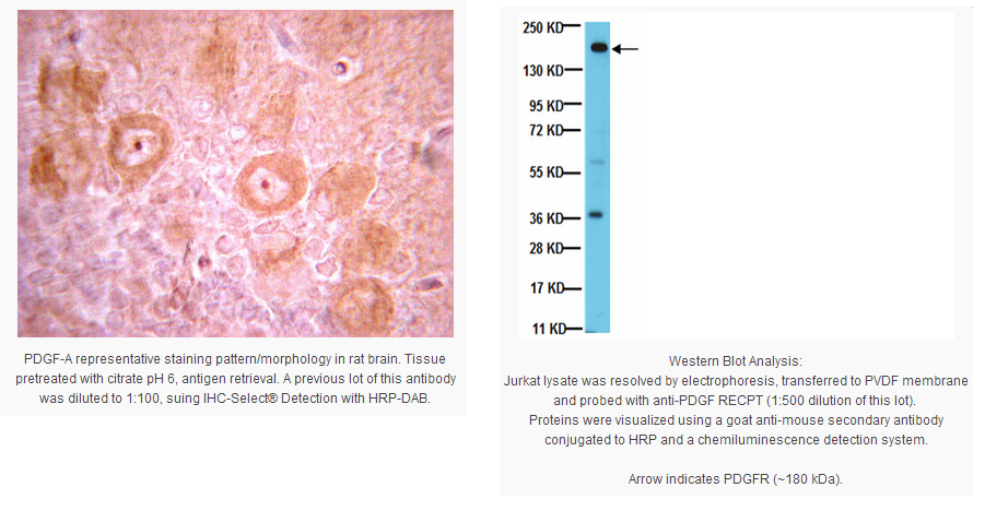
Immunohistochemistry(paraffin): Representative images from a previous lot.
Optimal Staining of PDGF-A With Citrate pH 6.0 Epitope Retrieval: Rat Brain
Routinely evaluated by Western Blot on Jurkat lysates.
Western Blot Analysis: 1:500 dilution of this lot detected PDGFR on 10 μg of Jurkat lysates.
Usage Statement
Unless otherwise stated in our catalog or other company documentation accompanying the product(s), our products are intended for research use only and are not to be used for any other purpose, which includes but is not limited to, unauthorized commercial uses, in vitro diagnostic uses, ex vivo or in vivo therapeutic uses or any type of consumption or application to humans or animals.
Storage and Shipping Information
Storage Conditions
Maintain at + 2-8 ° C in undiluted aliquots for up to 1 year from date of receipt.
Packaging Information
Material Size
100 µg
原厂资料:
Key Spec Table
Species Reactivity
Key Applications
Host
Format
Antibody Type
H, R, M
WB, IH(P), ELISA
M
Purified
Monoclonal Antibody
Description
Catalogue Number
05-1135
Replaces
04-397
Description
Anti-PDGFR Antibody, clone CH-3
Alternate Names
Platelet Derived Growth Factor Receptor Beta
CD140b
PDGFR-1
Platelet-derived growth factor receptor 1
CD140 antigen-like family member B
Beta-type platelet-derived growth factor receptor
Beta platelet-derived growth factor receptor
PDGFR-beta
PDGF-R-beta
Background Information
PDGFR (Platelet-Derived Growth Factor Receptor) is a receptor tyrosine kinase for the PDGF family of growth factors. PDGF homo and hetero- dimmers (dimer of A (17 kDa) and B (14 kDa) chains (AA, AB, or BB)) bind to the two structurally related a and b PDGF Receptors (PDGFRa and PDGFRb). The type A PDGF Receptor binds all three PDGFs with high affinity, whereas the type B PDGF Receptor is selective for PDGF-BB. This binding results in the dimerization of the receptor and the subsequent kinase activation of the receptor tyrosine kinase (RTK) and its autophosphorylation. These phosphorylations allow for various proteins, such as PI3 Kinase, PLCg, and Grb2, to bind through their SH2 domains and transduce signals into the cell. The activation of PDGFFR triggers the intracellular loop and activates Ras, Erk1/2, and ROS. PDGFb has been implicated in embryonic development, angiogenesis, and development of tumors of glial origin.
Product Information
Format
Purified
Control
Jurkat cell lysate
Presentation
Purified mouse monoclonal IgG1 in buffer containing 20 mM sodium phosphate, 250 mM NaCl, 0.1% NaN3, pH. 7.6
Applications
Application
This Anti-PDGFR Antibody, clone CH-3 is validated for use in WB, ELISA, IH(P) for the detection of PDGFR.
Key Applications
Western Blotting
Immunohistochemistry (Paraffin)
ELISA
Application Notes
ELISA: A previous lot of this antibody was used in ELISA.
Immunohistochemistry(paraffin): Representative images from a previous lot.
Optimal Staining of PDGF-A With Citrate pH 6.0 Epitope Retrieval: Rat Brain
Routinely evaluated by Western Blot on Jurkat lysates.
Western Blot Analysis: 1:500 dilution of this lot detected PDGFR on 10 μg of Jurkat lysates.
Usage Statement
Unless otherwise stated in our catalog or other company documentation accompanying the product(s), our products are intended for research use only and are not to be used for any other purpose, which includes but is not limited to, unauthorized commercial uses, in vitro diagnostic uses, ex vivo or in vivo therapeutic uses or any type of consumption or application to humans or animals.
Storage and Shipping Information
Storage Conditions
Maintain at + 2-8 ° C in undiluted aliquots for up to 1 year from date of receipt.


 京公网安备11010802025653 版权所有:北京逸优科技有限公司
