signal transducer and activator of transcription 1
Background Information
STATs (Signal-Transducing Activators of Transcription) are the principle substrates of JAK kinases, and mediators of cytokine signaling. STAT proteins have SH2 domains, and once phosphorylated by JAK kinases, STATs dimerize in a head-to-tail fashion, through their SH2 domains. STAT1 is a critical mediator of IFN signaling, STAT4 is specific to IL-12, STAT6 is activated primarily by IL-4 and IL-13, while STAT5 A and B appear to be most important to the function of growth hormone and prolactin. STAT3 is involved in multiple signaling pathways, and mice deficient in it are not viable. In addition to tyrosine phosphorylation, many STAT proteins are targets of Ser/Thr kinases, and those phosphorylation events potentiate STAT-induced transcription. The phosphorylation state of STATs can be monitored by phosphorylation state-specific antibodies.
Product Information
Format
Purified
Control
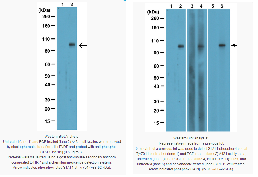
EGF treated and untreated A431 cell lysates, PDGF treated and untreated NIH/3T3 cell lysates, pervanadate treated and untreated PC12 cell lysates.
Presentation
Purified mouse monoclonal IgG1κ in buffer containing PBS and 0.05% sodium azide.
Applications
Application
Anti-Phospho-Stat1 (Tyr701) Antibody, clone 5C9.2 detects level of Phospho-Stat1 (Tyr701) & has been published & validated for use in WB.
Key Applications
Western Blotting
Biological Information
Immunogen
KLH-conjugated linear peptide corresponding to the region around phosphorylated tyrosine 701.
Epitope
Phosphorylated Tyr701
Clone
5C9.2
Concentration
Please refer to the Certificate of Analysis for the lot-specific concentration.
Host
Mouse
Specificity
This antibody recognizes phosphorylated STAT1 at tyrosine 701.
The protein encoded by this gene is a member of the STAT protein family. In response to cytokines and growth factors, STAT family members are phosphorylated by the receptor associated kinases, and then form homo- or heterodimers that translocate to the cell nucleus where they act as transcription activators. This protein can be activated by various ligands including interferon-alpha, interferon-gamma, EGF, PDGF and IL6. This protein mediates the expression of a variety of genes, which is thought to be important for cell viability in response to different cell stimuli and pathogens. Two alternatively spliced transcript variants encoding distinct isoforms have been described. [provided by RefSeq]
Function:Signal transducer and activator of transcription that mediates signaling by interferons (IFNs). Following type I IFN (IFN-alpha and IFN-beta) binding to cell surface receptors, Jak kinases (TYK2 and JAK1) are activated, leading to tyrosine phosphorylation of STAT1 and STAT2. The phosphorylated STATs dimerize, associate with ISGF3G/IRF-9 to form a complex termed ISGF3 transcription factor, that enters the nucleus. ISGF3 binds to the IFN stimulated response element (ISRE) to activate the transcription of interferon stimulated genes, which drive the cell in an antiviral state. In response to type II IFN (IFN-gamma), STAT1 is tyrosine- and serine-phosphorylated. It then forms a homodimer termed IFN-gamma-activated factor (GAF), migrates into the nucleus and binds to the IFN gamma activated sequence (GAS) to drive the expression of the target genes, inducing a cellular antiviral state.
Subunit structure:Isoform alpha homodimerizes upon IFN-gamma induced phosphorylation. Heterodimer with STAT2 upon IFN-alpha/beta induced phosphorylation. Interacts with NMI. Interacts with Sendai virus C', C, Y1 and Y2 proteins, Nipah virus P, V and W proteins, and rabies virus phosphoprotein preventing activation of ISRE and GAS promoter By similarity. Interaction with HCV core protein results in STAT1 degradation.
Subcellular location:Cytoplasm. Nucleus. Note= Translocated into the nucleus upon activation by IFN-alpha/beta.
Post-translational modification: Phosphorylated on tyrosine residues in response to IFN-alpha, IFN-gamma, PDGF and EGF. Serine phosphorylation is also required for maximal transcriptional activity in IFN-gamma transduction (lacking in beta form). Ref.6 Ref.7 Ref.11 Ref.12
Involvement in disease:Defects in STAT1 are the cause of STAT1 deficiency [MIM:600555]. Patients generally suffer from mycobacterial or viral diseases. In the case of complete deficiency, patients can die of viral disease. Ref.15
Defects in STAT1 are a cause of mendelian susceptibility to mycobacterial disease (MSMD) [MIM:209950]; also known as familial disseminated atypical mycobacterial infection. This rare condition confers predisposition to illness caused by moderately virulent mycobacterial species, such as Bacillus Calmette-Guerin (BCG) vaccine and environmental non-tuberculous mycobacteria, and by the more virulent Mycobacterium tuberculosis. Other microorganisms rarely cause severe clinical disease in individuals with susceptibility to mycobacterial infections, with the exception of Salmonella which infects less than 50% of these individuals. The pathogenic mechanism underlying MSMD is the impairment of interferon-gamma mediated immunity whose severity determines the clinical outcome. Some patients die of overwhelming mycobacterial disease with lepromatous-like lesions in early childhood, whereas others develop, later in life, disseminated but curable infections with tuberculoid granulomas. MSMD is a genetically heterogeneous disease with autosomal recessive, autosomal dominant or X-linked inheritance. Ref.14
Sequence similarities: Belongs to the transcription factor STAT family.
Contains 1 SH2 domain.
Product Usage Statements
Quality Assurance
Routinely evaluated by western blot on EGF treated and untreated A431 cell lysate.
Western Blot Analysis: 0.5-2 µg/mL of this lot was used to detect STAT1 phosphorylated at Tyr701 in EGF treated and untreated A431 cell lysates.
Usage Statement
Unless otherwise stated in our catalog or other company documentation accompanying the product(s), our products are intended for research use only and are not to be used for any other purpose, which includes but is not limited to, unauthorized commercial uses, in vitro diagnostic uses, ex vivo or in vivo therapeutic uses or any type of consumption or application to humans or animals.
Storage and Shipping Information
Storage Conditions
Stable for 1 year at 2-8ºC from date of receipt.
Packaging Information
Material Size
100 µg
原厂资料:
Key Spec Table
Species Reactivity
Key Applications
Host
Format
Antibody Type
H, R, M
WB
M
Purified
Monoclonal Antibody
Description
Catalogue Number
05-1064
Description
Anti-Phospho-Stat1 (Tyr701) Antibody, clone 5C9.2
Alternate Names
Transcription factor ISGF-3 components p91/p84
signal transducer and activator of transcription 1
Background Information
STATs (Signal-Transducing Activators of Transcription) are the principle substrates of JAK kinases, and mediators of cytokine signaling. STAT proteins have SH2 domains, and once phosphorylated by JAK kinases, STATs dimerize in a head-to-tail fashion, through their SH2 domains. STAT1 is a critical mediator of IFN signaling, STAT4 is specific to IL-12, STAT6 is activated primarily by IL-4 and IL-13, while STAT5 A and B appear to be most important to the function of growth hormone and prolactin. STAT3 is involved in multiple signaling pathways, and mice deficient in it are not viable. In addition to tyrosine phosphorylation, many STAT proteins are targets of Ser/Thr kinases, and those phosphorylation events potentiate STAT-induced transcription. The phosphorylation state of STATs can be monitored by phosphorylation state-specific antibodies.
Product Information
Format
Purified
Control
EGF treated and untreated A431 cell lysates, PDGF treated and untreated NIH/3T3 cell lysates, pervanadate treated and untreated PC12 cell lysates.
Presentation
Purified mouse monoclonal IgG1κ in buffer containing PBS and 0.05% sodium azide.
Applications
Application
Anti-Phospho-Stat1 (Tyr701) Antibody, clone 5C9.2 detects level of Phospho-Stat1 (Tyr701) & has been published & validated for use in WB.
Key Applications
Western Blotting
Biological Information
Immunogen
KLH-conjugated linear peptide corresponding to the region around phosphorylated tyrosine 701.
Epitope
Phosphorylated Tyr701
Clone
5C9.2
Concentration
Please refer to the Certificate of Analysis for the lot-specific concentration.
Host
Mouse
Specificity
This antibody recognizes phosphorylated STAT1 at tyrosine 701.
The protein encoded by this gene is a member of the STAT protein family. In response to cytokines and growth factors, STAT family members are phosphorylated by the receptor associated kinases, and then form homo- or heterodimers that translocate to the cell nucleus where they act as transcription activators. This protein can be activated by various ligands including interferon-alpha, interferon-gamma, EGF, PDGF and IL6. This protein mediates the expression of a variety of genes, which is thought to be important for cell viability in response to different cell stimuli and pathogens. Two alternatively spliced transcript variants encoding distinct isoforms have been described. [provided by RefSeq]
Function:Signal transducer and activator of transcription that mediates signaling by interferons (IFNs). Following type I IFN (IFN-alpha and IFN-beta) binding to cell surface receptors, Jak kinases (TYK2 and JAK1) are activated, leading to tyrosine phosphorylation of STAT1 and STAT2. The phosphorylated STATs dimerize, associate with ISGF3G/IRF-9 to form a complex termed ISGF3 transcription factor, that enters the nucleus. ISGF3 binds to the IFN stimulated response element (ISRE) to activate the transcription of interferon stimulated genes, which drive the cell in an antiviral state. In response to type II IFN (IFN-gamma), STAT1 is tyrosine- and serine-phosphorylated. It then forms a homodimer termed IFN-gamma-activated factor (GAF), migrates into the nucleus and binds to the IFN gamma activated sequence (GAS) to drive the expression of the target genes, inducing a cellular antiviral state.
Subunit structure:Isoform alpha homodimerizes upon IFN-gamma induced phosphorylation. Heterodimer with STAT2 upon IFN-alpha/beta induced phosphorylation. Interacts with NMI. Interacts with Sendai virus C', C, Y1 and Y2 proteins, Nipah virus P, V and W proteins, and rabies virus phosphoprotein preventing activation of ISRE and GAS promoter By similarity. Interaction with HCV core protein results in STAT1 degradation.
Subcellular location:Cytoplasm. Nucleus. Note= Translocated into the nucleus upon activation by IFN-alpha/beta.
Post-translational modification: Phosphorylated on tyrosine residues in response to IFN-alpha, IFN-gamma, PDGF and EGF. Serine phosphorylation is also required for maximal transcriptional activity in IFN-gamma transduction (lacking in beta form). Ref.6 Ref.7 Ref.11 Ref.12
Involvement in disease:Defects in STAT1 are the cause of STAT1 deficiency [MIM:600555]. Patients generally suffer from mycobacterial or viral diseases. In the case of complete deficiency, patients can die of viral disease. Ref.15
Defects in STAT1 are a cause of mendelian susceptibility to mycobacterial disease (MSMD) [MIM:209950]; also known as familial disseminated atypical mycobacterial infection. This rare condition confers predisposition to illness caused by moderately virulent mycobacterial species, such as Bacillus Calmette-Guerin (BCG) vaccine and environmental non-tuberculous mycobacteria, and by the more virulent Mycobacterium tuberculosis. Other microorganisms rarely cause severe clinical disease in individuals with susceptibility to mycobacterial infections, with the exception of Salmonella which infects less than 50% of these individuals. The pathogenic mechanism underlying MSMD is the impairment of interferon-gamma mediated immunity whose severity determines the clinical outcome. Some patients die of overwhelming mycobacterial disease with lepromatous-like lesions in early childhood, whereas others develop, later in life, disseminated but curable infections with tuberculoid granulomas. MSMD is a genetically heterogeneous disease with autosomal recessive, autosomal dominant or X-linked inheritance. Ref.14
Sequence similarities: Belongs to the transcription factor STAT family.
Contains 1 SH2 domain.
Product Usage Statements
Quality Assurance
Routinely evaluated by western blot on EGF treated and untreated A431 cell lysate.
Western Blot Analysis: 0.5-2 µg/mL of this lot was used to detect STAT1 phosphorylated at Tyr701 in EGF treated and untreated A431 cell lysates.
Usage Statement
Unless otherwise stated in our catalog or other company documentation accompanying the product(s), our products are intended for research use only and are not to be used for any other purpose, which includes but is not limited to, unauthorized commercial uses, in vitro diagnostic uses, ex vivo or in vivo therapeutic uses or any type of consumption or application to humans or animals.


 京公网安备11010802025653 版权所有:北京逸优科技有限公司
