Akt is the major known effector of the PI3 Kinase pathway. Generation of PIP3 results in the activation of PDK1, which phosphorylates Akt on Thr308, and another kinase complex, thought to be the mTORC2 complex (or possibly an intermediate kinase linking the two) that phosphorylates Akt on Ser473. These phosphorylations additively activate Akt Ser/Thr kinase activity, and the use of phosphorylation state-specific antibodies directed against either of these sites can imply Akt activation. Activation of Akt can be measured directly by immunoprecipitation followed by phosphorylation of a known substrate with radiolabeled ATP. Akt has been shown to phosphorylate over 70 substrates including TSC1&2 in the mTOR pathway, Bad on Ser136, GSK3, which is inactivated by this phosphorylation, the FoxO family of transcription factors, PRAS40, AS160.
This Anti-phospho-Akt1/PKBα (Thr308) Antibody, clone NL50 is validated for use in WB for the detection of phospho-Akt1/PKBα (Thr308).
Key Applications
Western Blotting
Application Notes
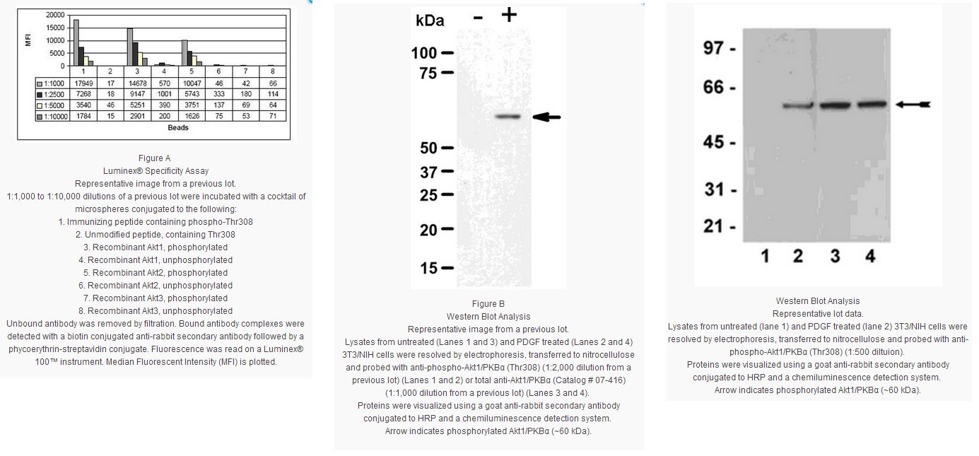
Immunoblot Analysis: A 1:500-1:2,000 dilution of this lot detected phospho-Akt1/PKBα in RIPA lysates from 3T3/NIH cells stimulated with 100ng/ml PDGF for 20 minutes (Figure A).
Beadlyte® Specificity Assay: 1:1,000-1:10,000 dilutions of this lot were incubated with phosphorylated and unphosphorylated recombinant Akt proteins conjugated to Luminex microspheres. Cross-reactivity to active Akt2 was detected (Figure B).
Biological Information
Immunogen
KLH-conjugated, synthetic peptide containing the sequence ..MK[pT]FC.. in which pT corresponds to phospho-threonine at residue 308 of mouse Akt. The Akt1 sequence is identical to human, rat, bovine and chicken Akt1. Human and mouse Akt2 shares 11/12
amino acids and rat Akt3 shares 10/12 amino acids with the peptide immunogen sequence.
Epitope
pThr308
Clone
NL50
Host
Rabbit
Specificity
Recognizes phosphorylated Akt1/PKBα, Mr 60 kDa. Cross-reactivity observed with Akt2, but not with Akt3.
Isotype
IgG
Species Reactivity
Rat Human Mouse Bovine Chicken
Species Reactivity Note
Human and mouse. Predicted to cross-react with rat, bovine and chicken based on sequence homology.
The serine-threonine protein kinase encoded by the AKT1 gene is catalytically inactive in serum-starved primary and immortalized fibroblasts. AKT1 and the related AKT2 are activated by platelet-derived growth factor. The activation is rapid and specific, and it is abrogated by mutations in the pleckstrin homology domain of AKT1. It was shown that the activation occurs through phosphatidylinositol 3-kinase. In the developing nervous system AKT is a critical mediator of growth factor-induced neuronal survival. Survival factors can suppress apoptosis in a transcription-independent manner by activating the serine/threonine kinase AKT1, which then phosphorylates and inactivates components of the apoptotic machinery. Multiple alternatively spliced transcript variants have been found for this gene.
FUNCTION:SwissProt: P31749 # General protein kinase capable of phosphorylating several known proteins. Phosphorylates TBC1D4. Signals downstream of phosphatidylinositol 3-kinase (PI(3)K) to mediate the effects of various growth factors such as platelet-derived growth factor (PDGF), epidermal growth factor (EGF), insulin and insulin-like growth factor I (IGF-I). Plays a role in glucose transport by mediating insulin-induced translocation of the GLUT4 glucose transporter to the cell surface. Mediates the antiapoptotic effects of IGF-I. Mediates insulin-stimulated protein synthesis, partly by playing a role in both insulin-induced phosphorylation of 4E-BP1 and in insulin-induced activation of p70 S6 kinase. Promotes glycogen synthesis by mediating the insulin-induced activation of glycogen synthase. SIZE:480 amino acids; 55686 Da SUBUNIT:Interacts with CENTG1 isoform 2 (PIKE-A) in the presence of guanine nucleotides. The C-terminus interacts with CCDC88A/GRDN. Interacts with AKTIP. SUBCELLULAR LOCATION:Cytoplasm. Nucleus. Note=Nucleus after activation by integrin-linked protein kinase 1 (ILK1). TISSUE SPECIFICITY:In all human cell types so far analyzed. DOMAIN:SwissProt: P31749 Binding of the PH domain to the phosphatidylinositol 3- kinase alpha (PI(3)K) results in its targeting to the plasma membrane. PTM:Phosphorylation on Thr-308, Ser-473 and Tyr-474 is required for full activity. Ser-473 phosphorylation by the Rictor-mTor complex favors Thr-308 phosphorylation by PDPK1. Ser-473 phosphorylation is enhanced by interaction with CENTG1 isoform 2 (PIKE-A). SIMILARITY:Belongs to the protein kinase superfamily. AGC Ser/Thr protein kinase family. RAC subfamily. & Contains 1 AGC-kinase C-terminal domain. & Contains 1 PH domain. & Contains 1 protein kinase domain.
Product Usage Statements
Quality Assurance
Evaluated by western blot on RIPA lysates from 3T3/NIH cells stimulated with 100 ng/mL PDGF for 20 minutes.
Western Blotting Analysis: A 1:500 dilution of this antibody detected phospho-Akt1/PKBα in RIPA lysates from 3T3/NIH cells stimulated with 100 ng/mL PDGF for 20 minutes.
Usage Statement
Unless otherwise stated in our catalog or other company documentation accompanying the product(s), our products are intended for research use only and are not to be used for any other purpose, which includes but is not limited to, unauthorized commercial uses, in vitro diagnostic uses, ex vivo or in vivo therapeutic uses or any type of consumption or application to humans or animals.
Storage and Shipping Information
Storage Conditions
Stable for 1 year at -20°C from date of receipt.
Handling Recommendations: Upon receipt, and prior to removing the cap, centrifuge the vial and gently mix the solution. Aliquot into microcentrifuge tubes and store at -20°C. Avoid repeated freeze/thaw cycles, which may damage IgG and affect product performance.
Akt is the major known effector of the PI3 Kinase pathway. Generation of PIP3 results in the activation of PDK1, which phosphorylates Akt on Thr308, and another kinase complex, thought to be the mTORC2 complex (or possibly an intermediate kinase linking the two) that phosphorylates Akt on Ser473. These phosphorylations additively activate Akt Ser/Thr kinase activity, and the use of phosphorylation state-specific antibodies directed against either of these sites can imply Akt activation. Activation of Akt can be measured directly by immunoprecipitation followed by phosphorylation of a known substrate with radiolabeled ATP. Akt has been shown to phosphorylate over 70 substrates including TSC1&2 in the mTOR pathway, Bad on Ser136, GSK3, which is inactivated by this phosphorylation, the FoxO family of transcription factors, PRAS40, AS160.
This Anti-phospho-Akt1/PKBα (Thr308) Antibody, clone NL50 is validated for use in WB for the detection of phospho-Akt1/PKBα (Thr308).
Key Applications
Western Blotting
Application Notes
Immunoblot Analysis: A 1:500-1:2,000 dilution of this lot detected phospho-Akt1/PKBα in RIPA lysates from 3T3/NIH cells stimulated with 100ng/ml PDGF for 20 minutes (Figure A).
Beadlyte® Specificity Assay: 1:1,000-1:10,000 dilutions of this lot were incubated with phosphorylated and unphosphorylated recombinant Akt proteins conjugated to Luminex microspheres. Cross-reactivity to active Akt2 was detected (Figure B).
Biological Information
Immunogen
KLH-conjugated, synthetic peptide containing the sequence ..MK[pT]FC.. in which pT corresponds to phospho-threonine at residue 308 of mouse Akt. The Akt1 sequence is identical to human, rat, bovine and chicken Akt1. Human and mouse Akt2 shares 11/12
amino acids and rat Akt3 shares 10/12 amino acids with the peptide immunogen sequence.
Epitope
pThr308
Clone
NL50
Host
Rabbit
Specificity
Recognizes phosphorylated Akt1/PKBα, Mr 60 kDa. Cross-reactivity observed with Akt2, but not with Akt3.
Isotype
IgG
Species Reactivity
Rat Human Mouse Bovine Chicken
Species Reactivity Note
Human and mouse. Predicted to cross-react with rat, bovine and chicken based on sequence homology.
The serine-threonine protein kinase encoded by the AKT1 gene is catalytically inactive in serum-starved primary and immortalized fibroblasts. AKT1 and the related AKT2 are activated by platelet-derived growth factor. The activation is rapid and specific, and it is abrogated by mutations in the pleckstrin homology domain of AKT1. It was shown that the activation occurs through phosphatidylinositol 3-kinase. In the developing nervous system AKT is a critical mediator of growth factor-induced neuronal survival. Survival factors can suppress apoptosis in a transcription-independent manner by activating the serine/threonine kinase AKT1, which then phosphorylates and inactivates components of the apoptotic machinery. Multiple alternatively spliced transcript variants have been found for this gene.
FUNCTION:SwissProt: P31749 # General protein kinase capable of phosphorylating several known proteins. Phosphorylates TBC1D4. Signals downstream of phosphatidylinositol 3-kinase (PI(3)K) to mediate the effects of various growth factors such as platelet-derived growth factor (PDGF), epidermal growth factor (EGF), insulin and insulin-like growth factor I (IGF-I). Plays a role in glucose transport by mediating insulin-induced translocation of the GLUT4 glucose transporter to the cell surface. Mediates the antiapoptotic effects of IGF-I. Mediates insulin-stimulated protein synthesis, partly by playing a role in both insulin-induced phosphorylation of 4E-BP1 and in insulin-induced activation of p70 S6 kinase. Promotes glycogen synthesis by mediating the insulin-induced activation of glycogen synthase. SIZE:480 amino acids; 55686 Da SUBUNIT:Interacts with CENTG1 isoform 2 (PIKE-A) in the presence of guanine nucleotides. The C-terminus interacts with CCDC88A/GRDN. Interacts with AKTIP. SUBCELLULAR LOCATION:Cytoplasm. Nucleus. Note=Nucleus after activation by integrin-linked protein kinase 1 (ILK1). TISSUE SPECIFICITY:In all human cell types so far analyzed. DOMAIN:SwissProt: P31749 Binding of the PH domain to the phosphatidylinositol 3- kinase alpha (PI(3)K) results in its targeting to the plasma membrane. PTM:Phosphorylation on Thr-308, Ser-473 and Tyr-474 is required for full activity. Ser-473 phosphorylation by the Rictor-mTor complex favors Thr-308 phosphorylation by PDPK1. Ser-473 phosphorylation is enhanced by interaction with CENTG1 isoform 2 (PIKE-A). SIMILARITY:Belongs to the protein kinase superfamily. AGC Ser/Thr protein kinase family. RAC subfamily. & Contains 1 AGC-kinase C-terminal domain. & Contains 1 PH domain. & Contains 1 protein kinase domain.
Product Usage Statements
Quality Assurance
Evaluated by western blot on RIPA lysates from 3T3/NIH cells stimulated with 100 ng/mL PDGF for 20 minutes.
Western Blotting Analysis: A 1:500 dilution of this antibody detected phospho-Akt1/PKBα in RIPA lysates from 3T3/NIH cells stimulated with 100 ng/mL PDGF for 20 minutes.
Usage Statement
Unless otherwise stated in our catalog or other company documentation accompanying the product(s), our products are intended for research use only and are not to be used for any other purpose, which includes but is not limited to, unauthorized commercial uses, in vitro diagnostic uses, ex vivo or in vivo therapeutic uses or any type of consumption or application to humans or animals.
Storage and Shipping Information
Storage Conditions
Stable for 1 year at -20°C from date of receipt.
Handling Recommendations: Upon receipt, and prior to removing the cap, centrifuge the vial and gently mix the solution. Aliquot into microcentrifuge tubes and store at -20°C. Avoid repeated freeze/thaw cycles, which may damage IgG and affect product performance.


 京公网安备11010802025653 版权所有:北京逸优科技有限公司
