Human platelet-derived growth factor (PDGF) receptor, is a receptor tyrosine kinase whose activity induces various signalling cascades leading to cell growth and proliferation. Two major isoforms exist, PDGF receptor-Alpha and PDGF receptor-Beta. PDGF receptor-Beta can only bind two specific PDGF isoforms, PDGF-Beta and PDGFDelta (1,2). Upon ligand binding, the receptor undergoes autophosphorylation, allowing binding and activation of cytoplasmic SH2 domain-containing signal transduction molecules such as PI kinase (3), Grb2, and Src, among others.
Product Information
Format
Unpurified
Control
NIH/3T3 cell lysate
Presentation
Rabbit Monoclonal in buffer containing glycerol, BSA, and sodium azide.
Applications
Application
Please note that this product will not be available for sale after March 15, 2015. Please select one of the other antibodies against this target.
Key Applications
Immunohistochemistry (Paraffin)
Western Blotting
Immunocytochemistry
Immunoprecipitation
Application Notes
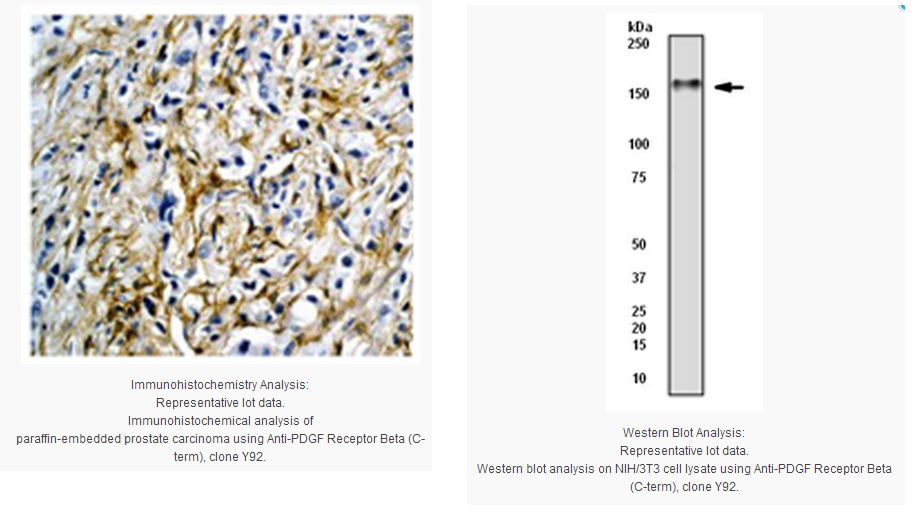
Immunohistochemistry Analysis: A 1:100 dilution from a representative lot was used in prostate carcinoma tissue.
Immunocytochemistry Analysis: A 1:100 dilution from a representative lot was used in IC.
Immunoprecipitation Analysis: A 1:30 dilution from a representative lot was used in IP.
Biological Information
Immunogen
Synthetic peptide corresponding to the C-terminus of human PDGF Receptor Beta.
Epitope
C-terminus
Clone
Y92
Host
Rabbit
Specificity
This antibody recognizes the C-terminus of PDGF Receptor Beta. The antibody does not cross-react with other CSF-1/PDGF receptor family members.
FUNCTION:Tyrosine-protein kinase that acts as cell-surface receptor for homodimeric PDGFB and PDGFD and for heterodimers formed by PDGFA and PDGFB, and plays an essential role in the regulation of embryonic development, cell proliferation, survival, differentiation, chemotaxis and migration. Plays an essential role in blood vessel development by promoting proliferation, migration and recruitment of pericytes and smooth muscle cells to endothelial cells. Plays a role in the migration of vascular smooth muscle cells and the formation of neointima at vascular injury sites. Required for normal development of the cardiovascular system. Required for normal recruitment of pericytes (mesangial cells) in the kidney glomerulus, and for normal formation of a branched network of capillaries in kidney glomeruli. Promotes rearrangement of the actin cytoskeleton and the formation of membrane ruffles. Binding of its cognate ligands - homodimeric PDGFB, heterodimers formed by PDGFA and PDGFB or homodimeric PDGFD -leads to the activation of several signaling cascades; the response depends on the nature of the bound ligand and is modulated by the formation of heterodimers between PDGFRA and PDGFRB. Phosphorylates PLCG1, PIK3R1, PTPN11, RASA1/GAP, CBL, SHC1 and NCK1. Activation of PLCG1 leads to the production of the cellular signaling molecules diacylglycerol and inositol-1,4,5-trisphosphate, mobilization of cytosolic Ca2+ and the activation of protein kinase C. Phosphorylation of PIK3R1, the regulatory subunit of phosphatidylinositol 3-kinase, leads to the activation of the AKT1 signaling pathway. Phosphorylation of SHC1, or of the C-terminus of PTPN11, creates a binding site for GRB2, resulting in the activation of HRAS, RAF1 and down-stream MAP kinases, including MAPK1/ERK2 and/or MAPK3/ERK1. Promotes phosphorylation and activation of SRC family kinases. Promotes phosphorylation of PDCD6IP/ALIX and STAM. Receptor signaling is down-regulated by protein phosphatases that dephosphorylate the receptor and its down-stream effectors, and by rapid internalization of the activated receptor. CATALYTIC ACTIVITY:ATP + a [protein]-L-tyrosine = ADP + a [protein]-L-tyrosine phosphate. ENZYME REGULATION:Present in an inactive conformation in the absence of bound ligand. Binding of PDGFB and/or PDGFD leads to dimerization and activation by autophosphorylation on tyrosine residues. Inhibited by imatinib. SUBUNIT STRUCTURE:Interacts with homodimeric PDGFB and PDGFD, and with heterodimers formed by PDGFA and PDGFB. May also interact with homodimeric PDGFC. Monomer in the absence of bound ligand. Interaction with homodimeric PDGFB, heterodimers formed by PDGFA and PDGFB or homodimeric PDGFD, leads to receptor dimerization, where both PDGFRA homodimers and heterodimers with PDGFRB are observed. Interacts with SH2B2/APS. Interacts directly (tyrosine phosphorylated) with SHB. Interacts (tyrosine phosphorylated) with PIK3R1. Interacts (tyrosine phosphorylated) with CBL. Interacts (tyrosine phosphorylated) with SRC and SRC family kinases. Interacts (tyrosine phosphorylated) with PIK3C2B, maybe indirectly. Interacts (tyrosine phosphorylated) with SHC1, GRB7, GRB10 and NCK1. Interaction with GRB2 is mediated by SHC1. Interacts (via C-terminus) with SLC9A3R1. SUBCELLULAR LOCATION:Cell membrane; Single-pass type I membrane protein. Cytoplasmic vesicle. Lysosome lumen. Note: After ligand binding, the autophosphorylated receptor is ubiquitinated and internalized, leading to its degradation. PTM:Autophosphorylated on tyrosine residues upon ligand binding. Autophosphorylation occurs in trans, i.e. one subunit of the dimeric receptor phosphorylates tyrosine residues on the other subunit. Phosphorylation at Tyr-579, and to a lesser degree, at Tyr-581, is important for interaction with SRC family kinases. Phosphorylation at Tyr-740 and Tyr-751 is important for interaction with PIK3R1. Phosphorylation at Tyr-751 is important for interaction with NCK1. Phosphorylation at Tyr-771 and Tyr-857 is important for interaction with RASA1/GAP. Phosphorylation at Tyr-857 is important for efficient phosphorylation of PLCG1 and PTPN11, resulting in increased phosphorylation of AKT1, MAPK1/ERK2 and/or MAPK3/ERK1, PDCD6IP/ALIX and STAM, and in increased cell proliferation. Phosphorylation at Tyr-1009 is important for interaction with PTPN11. Phosphorylation at Tyr-1009 and Tyr-1021 is important for interaction with PLCG1. Phosphorylation at Tyr-1021 is important for interaction with CBL; PLCG1 and CBL compete for the same binding site. Dephosphorylated by PTPRJ at Tyr-751, Tyr-857, Tyr-1009 and Tyr-1021.
N-glycosylated. Ref.2 Ref.49
Ubiquitinated. After autophosphorylation, the receptor is polyubiquitinated, leading to its degradation. INVOLVEMENT IN DISEASE:Note=A chromosomal aberration involving PDGFRB is found in a form of chronic myelomonocytic leukemia (CMML). Translocation t(5;12)(q33;p13) with EVT6/TEL. It is characterized by abnormal clonal myeloid proliferation and by progression to acute myelogenous leukemia (AML).
Note=A chromosomal aberration involving PDGFRB may be a cause of acute myelogenous leukemia. Translocation t(5;14)(q33;q32) with TRIP11. The fusion protein may be involved in clonal evolution of leukemia and eosinophilia.
Note=A chromosomal aberration involving PDGFRB may be a cause of juvenile myelomonocytic leukemia. Translocation t(5;17)(q33;p11.2) with SPECC1.
Defects in PDGFRB are a cause of myeloproliferative disorder chronic with eosinophilia (MPE) [MIM:131440]. A hematologic disorder characterized by malignant eosinophils proliferation. Note=A chromosomal aberration involving PDGFRB is found in many instances of myeloproliferative disorder chronic with eosinophilia. Translocation t(5;12) with ETV6 on chromosome 12 creating an PDGFRB-ETV6 fusion protein. Translocation t(5;15)(q33;q22) with TP53BP1 creating a PDGFRB-TP53BP1 fusion protein.
Note=A chromosomal aberration involving PDGFRB may be the cause of a myeloproliferative disorder (MBD) associated with eosinophilia. Translocation t(1;5)(q23;q33) that forms a PDE4DIP-PDGFRB fusion protein.
Note=A chromosomal aberration involving PGFRB is found in a patient with T-lymphoblastic lymphoma (T-ALL) and an associated myeloproliferative neoplasm (MPN) with eosinophilia. Translocation t(5;6)(q33-34;q23) with CEP85L. The translocation fuses the 5'-end of CEP85L (isoform 4) to the 3'-end of PDGFRB. SEQUENCE SIMILARITIES:Belongs to the protein kinase superfamily. Tyr protein kinase family. CSF-1/PDGF receptor subfamily.
Contains 5 Ig-like C2-type (immunoglobulin-like) domains.
Contains 1 protein kinase domain.
Product Usage Statements
Quality Assurance
Evaluated by Western Blot in NIH/3T3 cell lysate
WesternBlot Analysis: A 1:10,000 dilution of this antibody detected PDGF Receptor Beta in NIH/3T3 cell lysate.
Usage Statement
Unless otherwise stated in our catalog or other company documentation accompanying the product(s), our products are intended for research use only and are not to be used for any other purpose, which includes but is not limited to, unauthorized commercial uses, in vitro diagnostic uses, ex vivo or in vivo therapeutic uses or any type of consumption or application to humans or animals.
Storage and Shipping Information
Storage Conditions
Stable for 1 year at -20ºC from date of receipt.
Handling Recommendations: Upon first thaw, and prior to removing cap, centrifuge the vial and gently mix the solution. Aliquot into microcentrifuge tubes and store at -20ºC. Avoid freeze/thaw cycles, which may damage IgG and affect product performance.
Note: Variability in freezer temperatures below -20°C may cause glycerol containing solutions to become frozen during storage. Note: Variability in freezer temperatures below -20°C may cause glycerol containing solutions to become frozen during storage.
Human platelet-derived growth factor (PDGF) receptor, is a receptor tyrosine kinase whose activity induces various signalling cascades leading to cell growth and proliferation. Two major isoforms exist, PDGF receptor-Alpha and PDGF receptor-Beta. PDGF receptor-Beta can only bind two specific PDGF isoforms, PDGF-Beta and PDGFDelta (1,2). Upon ligand binding, the receptor undergoes autophosphorylation, allowing binding and activation of cytoplasmic SH2 domain-containing signal transduction molecules such as PI kinase (3), Grb2, and Src, among others.
Product Information
Format
Unpurified
Control
NIH/3T3 cell lysate
Presentation
Rabbit Monoclonal in buffer containing glycerol, BSA, and sodium azide.
Applications
Application
Please note that this product will not be available for sale after March 15, 2015. Please select one of the other antibodies against this target.
Key Applications
Immunohistochemistry (Paraffin)
Western Blotting
Immunocytochemistry
Immunoprecipitation
Application Notes
Immunohistochemistry Analysis: A 1:100 dilution from a representative lot was used in prostate carcinoma tissue.
Immunocytochemistry Analysis: A 1:100 dilution from a representative lot was used in IC.
Immunoprecipitation Analysis: A 1:30 dilution from a representative lot was used in IP.
Biological Information
Immunogen
Synthetic peptide corresponding to the C-terminus of human PDGF Receptor Beta.
Epitope
C-terminus
Clone
Y92
Host
Rabbit
Specificity
This antibody recognizes the C-terminus of PDGF Receptor Beta. The antibody does not cross-react with other CSF-1/PDGF receptor family members.
FUNCTION:Tyrosine-protein kinase that acts as cell-surface receptor for homodimeric PDGFB and PDGFD and for heterodimers formed by PDGFA and PDGFB, and plays an essential role in the regulation of embryonic development, cell proliferation, survival, differentiation, chemotaxis and migration. Plays an essential role in blood vessel development by promoting proliferation, migration and recruitment of pericytes and smooth muscle cells to endothelial cells. Plays a role in the migration of vascular smooth muscle cells and the formation of neointima at vascular injury sites. Required for normal development of the cardiovascular system. Required for normal recruitment of pericytes (mesangial cells) in the kidney glomerulus, and for normal formation of a branched network of capillaries in kidney glomeruli. Promotes rearrangement of the actin cytoskeleton and the formation of membrane ruffles. Binding of its cognate ligands - homodimeric PDGFB, heterodimers formed by PDGFA and PDGFB or homodimeric PDGFD -leads to the activation of several signaling cascades; the response depends on the nature of the bound ligand and is modulated by the formation of heterodimers between PDGFRA and PDGFRB. Phosphorylates PLCG1, PIK3R1, PTPN11, RASA1/GAP, CBL, SHC1 and NCK1. Activation of PLCG1 leads to the production of the cellular signaling molecules diacylglycerol and inositol-1,4,5-trisphosphate, mobilization of cytosolic Ca2+ and the activation of protein kinase C. Phosphorylation of PIK3R1, the regulatory subunit of phosphatidylinositol 3-kinase, leads to the activation of the AKT1 signaling pathway. Phosphorylation of SHC1, or of the C-terminus of PTPN11, creates a binding site for GRB2, resulting in the activation of HRAS, RAF1 and down-stream MAP kinases, including MAPK1/ERK2 and/or MAPK3/ERK1. Promotes phosphorylation and activation of SRC family kinases. Promotes phosphorylation of PDCD6IP/ALIX and STAM. Receptor signaling is down-regulated by protein phosphatases that dephosphorylate the receptor and its down-stream effectors, and by rapid internalization of the activated receptor. CATALYTIC ACTIVITY:ATP + a [protein]-L-tyrosine = ADP + a [protein]-L-tyrosine phosphate. ENZYME REGULATION:Present in an inactive conformation in the absence of bound ligand. Binding of PDGFB and/or PDGFD leads to dimerization and activation by autophosphorylation on tyrosine residues. Inhibited by imatinib. SUBUNIT STRUCTURE:Interacts with homodimeric PDGFB and PDGFD, and with heterodimers formed by PDGFA and PDGFB. May also interact with homodimeric PDGFC. Monomer in the absence of bound ligand. Interaction with homodimeric PDGFB, heterodimers formed by PDGFA and PDGFB or homodimeric PDGFD, leads to receptor dimerization, where both PDGFRA homodimers and heterodimers with PDGFRB are observed. Interacts with SH2B2/APS. Interacts directly (tyrosine phosphorylated) with SHB. Interacts (tyrosine phosphorylated) with PIK3R1. Interacts (tyrosine phosphorylated) with CBL. Interacts (tyrosine phosphorylated) with SRC and SRC family kinases. Interacts (tyrosine phosphorylated) with PIK3C2B, maybe indirectly. Interacts (tyrosine phosphorylated) with SHC1, GRB7, GRB10 and NCK1. Interaction with GRB2 is mediated by SHC1. Interacts (via C-terminus) with SLC9A3R1. SUBCELLULAR LOCATION:Cell membrane; Single-pass type I membrane protein. Cytoplasmic vesicle. Lysosome lumen. Note: After ligand binding, the autophosphorylated receptor is ubiquitinated and internalized, leading to its degradation. PTM:Autophosphorylated on tyrosine residues upon ligand binding. Autophosphorylation occurs in trans, i.e. one subunit of the dimeric receptor phosphorylates tyrosine residues on the other subunit. Phosphorylation at Tyr-579, and to a lesser degree, at Tyr-581, is important for interaction with SRC family kinases. Phosphorylation at Tyr-740 and Tyr-751 is important for interaction with PIK3R1. Phosphorylation at Tyr-751 is important for interaction with NCK1. Phosphorylation at Tyr-771 and Tyr-857 is important for interaction with RASA1/GAP. Phosphorylation at Tyr-857 is important for efficient phosphorylation of PLCG1 and PTPN11, resulting in increased phosphorylation of AKT1, MAPK1/ERK2 and/or MAPK3/ERK1, PDCD6IP/ALIX and STAM, and in increased cell proliferation. Phosphorylation at Tyr-1009 is important for interaction with PTPN11. Phosphorylation at Tyr-1009 and Tyr-1021 is important for interaction with PLCG1. Phosphorylation at Tyr-1021 is important for interaction with CBL; PLCG1 and CBL compete for the same binding site. Dephosphorylated by PTPRJ at Tyr-751, Tyr-857, Tyr-1009 and Tyr-1021.
N-glycosylated. Ref.2 Ref.49
Ubiquitinated. After autophosphorylation, the receptor is polyubiquitinated, leading to its degradation. INVOLVEMENT IN DISEASE:Note=A chromosomal aberration involving PDGFRB is found in a form of chronic myelomonocytic leukemia (CMML). Translocation t(5;12)(q33;p13) with EVT6/TEL. It is characterized by abnormal clonal myeloid proliferation and by progression to acute myelogenous leukemia (AML).
Note=A chromosomal aberration involving PDGFRB may be a cause of acute myelogenous leukemia. Translocation t(5;14)(q33;q32) with TRIP11. The fusion protein may be involved in clonal evolution of leukemia and eosinophilia.
Note=A chromosomal aberration involving PDGFRB may be a cause of juvenile myelomonocytic leukemia. Translocation t(5;17)(q33;p11.2) with SPECC1.
Defects in PDGFRB are a cause of myeloproliferative disorder chronic with eosinophilia (MPE) [MIM:131440]. A hematologic disorder characterized by malignant eosinophils proliferation. Note=A chromosomal aberration involving PDGFRB is found in many instances of myeloproliferative disorder chronic with eosinophilia. Translocation t(5;12) with ETV6 on chromosome 12 creating an PDGFRB-ETV6 fusion protein. Translocation t(5;15)(q33;q22) with TP53BP1 creating a PDGFRB-TP53BP1 fusion protein.
Note=A chromosomal aberration involving PDGFRB may be the cause of a myeloproliferative disorder (MBD) associated with eosinophilia. Translocation t(1;5)(q23;q33) that forms a PDE4DIP-PDGFRB fusion protein.
Note=A chromosomal aberration involving PGFRB is found in a patient with T-lymphoblastic lymphoma (T-ALL) and an associated myeloproliferative neoplasm (MPN) with eosinophilia. Translocation t(5;6)(q33-34;q23) with CEP85L. The translocation fuses the 5'-end of CEP85L (isoform 4) to the 3'-end of PDGFRB. SEQUENCE SIMILARITIES:Belongs to the protein kinase superfamily. Tyr protein kinase family. CSF-1/PDGF receptor subfamily.
Contains 5 Ig-like C2-type (immunoglobulin-like) domains.
Contains 1 protein kinase domain.
Product Usage Statements
Quality Assurance
Evaluated by Western Blot in NIH/3T3 cell lysate
WesternBlot Analysis: A 1:10,000 dilution of this antibody detected PDGF Receptor Beta in NIH/3T3 cell lysate.
Usage Statement
Unless otherwise stated in our catalog or other company documentation accompanying the product(s), our products are intended for research use only and are not to be used for any other purpose, which includes but is not limited to, unauthorized commercial uses, in vitro diagnostic uses, ex vivo or in vivo therapeutic uses or any type of consumption or application to humans or animals.
Storage and Shipping Information
Storage Conditions
Stable for 1 year at -20ºC from date of receipt.
Handling Recommendations: Upon first thaw, and prior to removing cap, centrifuge the vial and gently mix the solution. Aliquot into microcentrifuge tubes and store at -20ºC. Avoid freeze/thaw cycles, which may damage IgG and affect product performance.
Note: Variability in freezer temperatures below -20°C may cause glycerol containing solutions to become frozen during storage. Note: Variability in freezer temperatures below -20°C may cause glycerol containing solutions to become frozen during storage.


 京公网安备11010802025653 版权所有:北京逸优科技有限公司
