Actin is a 43kDa highly conserved structural protein. There are three main actin isotypes (alpha, beta and gamma) which show > 90% amino-acid (aa) homology between isotypes and > 98% homology within members of a particular isotypic group. The majority of the isotype heterogeneity is located in the amino-terminal 30 aa. The different actin isotypes have been shown to behave very differently in vitro and in vivo. Recent studies describe differential subcellular localization of gamma-actin and isotype specific binding of actin associated proteins, (Pardo, 1983, Otey, 1986, Bassell, 1998).
Beta actin is one of the two nonmuscle cytoskeletal actins and is relatively stable and appears to remain at normal levels regardless of experimental treatment. It is generally used as an internal control for experiments.
Product Information
Format
Unpurified
Control
T47D and HeLa cell lysates.
Presentation
Rabbit Monoclonal in buffer containing 50mM Tris-Glycine (pH 7.4), 0.15M NaCl containing 40% Glycerol, 0.01% sodium azide and 0.05% BSA.
Applications
Application
Anti-beta Actin Antibody, clone EP1123Y, Rabbit detects level of beta Actin & has been published & validated for use in IH(P), WB & IC.
Key Applications
Immunocytochemistry
Immunohistochemistry (Paraffin)
Western Blotting
Application Notes
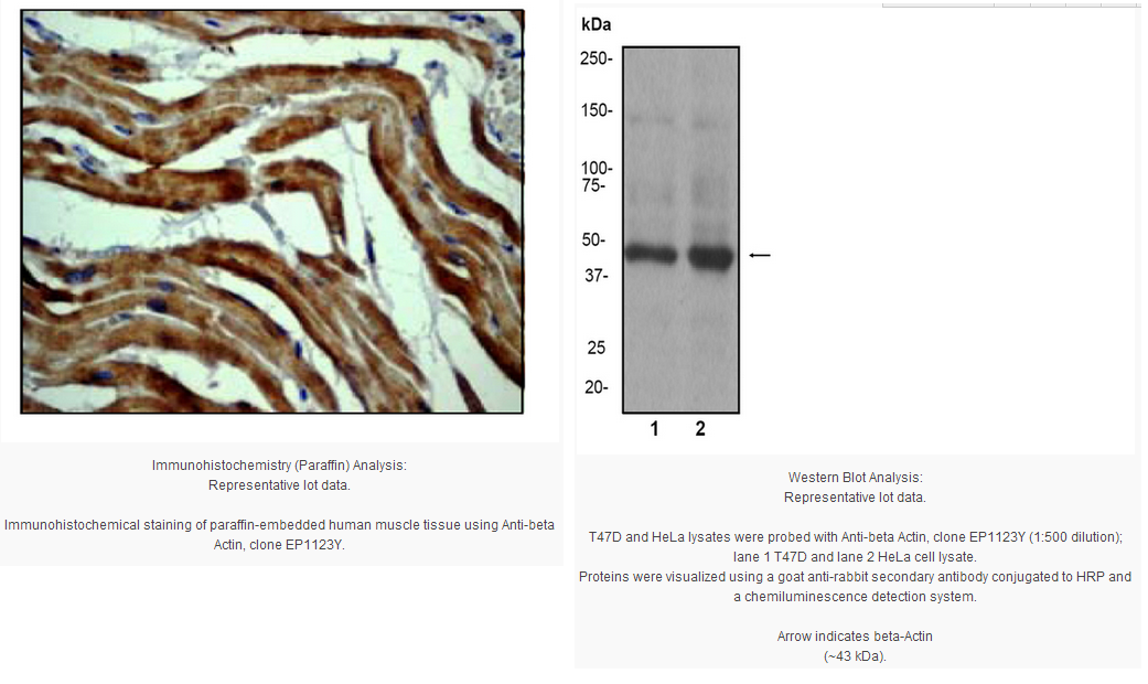
Immunohistochemistry Analysis: 1:50 dilution from a previous lot detected beta Actin in human muscle tissue.
Immunocytochemistry: 1:50 dilution from a previous lot was used on IC.
Biological Information
Immunogen
Synthetic peptide corresponding to residues near the C-terminus of human beta Actin.
FUNCTION:Actins are highly conserved proteins that are involved in various types of cell motility and are ubiquitously expressed in all eukaryotic cells. SUBUNIT STRUCTURE:Polymerization of globular actin (G-actin) leads to a structural filament (F-actin) in the form of a two-stranded helix. Each actin can bind to 4 others. Component of the BAF complex, which includes at least actin (ACTB), ARID1A, ARID1B/BAF250, SMARCA2, SMARCA4/BRG1, ACTL6A/BAF53, ACTL6B/BAF53B, SMARCE1/BAF57 SMARCC1/BAF155, SMARCC2/BAF170, SMARCB1/SNF5/INI1, and one or more of SMARCD1/BAF60A, SMARCD2/BAF60B, or SMARCD3/BAF60C. In muscle cells, the BAF complex also contains DPF3. Found in a complex with XPO6, Ran, ACTB and PFN1. Component of the MLL5-L complex, at least composed of MLL5, STK38, PPP1CA, PPP1CB, PPP1CC, HCFC1, ACTB and OGT. Interacts with XPO6. SUBCELLULAR LOCATION:Cytoplasm › cytoskeleton. INVOLVEMENT IN DISEASE:Defects in ACTB are a cause of dystonia juvenile-onset (DYTJ) [MIM:607371]. DYTJ is a form of dystonia with juvenile onset. Dystonia is defined by the presence of sustained involuntary muscle contraction, often leading to abnormal postures. DYTJ patients manifest progressive, generalized, dopa-unresponsive dystonia, developmental malformations and sensory hearing loss. MISCELLANEOUS:In vertebrates 3 main groups of actin isoforms, alpha, beta and gamma have been identified. The alpha actins are found in muscle tissues and are a major constituent of the contractile apparatus. The beta and gamma actins coexist in most cell types as components of the cytoskeleton and as mediators of internal cell motility. SEQUENCE SIMILARITIES:Belongs to the actin family.
Product Usage Statements
Quality Assurance
Evaluated by Western Blot on T47D and HeLa cell lysates.
Western Blot Analysis: 1:500 dilution of this antibody was used to detect beta Actin in T47D and HeLa cell lysates.
Usage Statement
Unless otherwise stated in our catalog or other company documentation accompanying the product(s), our products are intended for research use only and are not to be used for any other purpose, which includes but is not limited to, unauthorized commercial uses, in vitro diagnostic uses, ex vivo or in vivo therapeutic uses or any type of consumption or application to humans or animals.
Storage and Shipping Information
Storage Conditions
Stable for 1 year at -20ºC from date of receipt.
Handling Recommendations: Upon receipt, and prior to removing the cap, centrifuge the vial and gently mix the solution. Aliquot into microcentrifuge tubes and store at -20°C. Avoid repeated freeze/thaw cycles, which may damage IgG and affect product performance. Note: Variability in freezer temperatures below -20°C may cause glycerol containing solutions to become frozen during storage.
Actin is a 43kDa highly conserved structural protein. There are three main actin isotypes (alpha, beta and gamma) which show > 90% amino-acid (aa) homology between isotypes and > 98% homology within members of a particular isotypic group. The majority of the isotype heterogeneity is located in the amino-terminal 30 aa. The different actin isotypes have been shown to behave very differently in vitro and in vivo. Recent studies describe differential subcellular localization of gamma-actin and isotype specific binding of actin associated proteins, (Pardo, 1983, Otey, 1986, Bassell, 1998).
Beta actin is one of the two nonmuscle cytoskeletal actins and is relatively stable and appears to remain at normal levels regardless of experimental treatment. It is generally used as an internal control for experiments.
Product Information
Format
Unpurified
Control
T47D and HeLa cell lysates.
Presentation
Rabbit Monoclonal in buffer containing 50mM Tris-Glycine (pH 7.4), 0.15M NaCl containing 40% Glycerol, 0.01% sodium azide and 0.05% BSA.
Applications
Application
Anti-beta Actin Antibody, clone EP1123Y, Rabbit detects level of beta Actin & has been published & validated for use in IH(P), WB & IC.
Key Applications
Immunocytochemistry
Immunohistochemistry (Paraffin)
Western Blotting
Application Notes
Immunohistochemistry Analysis: 1:50 dilution from a previous lot detected beta Actin in human muscle tissue.
Immunocytochemistry: 1:50 dilution from a previous lot was used on IC.
Biological Information
Immunogen
Synthetic peptide corresponding to residues near the C-terminus of human beta Actin.
FUNCTION:Actins are highly conserved proteins that are involved in various types of cell motility and are ubiquitously expressed in all eukaryotic cells. SUBUNIT STRUCTURE:Polymerization of globular actin (G-actin) leads to a structural filament (F-actin) in the form of a two-stranded helix. Each actin can bind to 4 others. Component of the BAF complex, which includes at least actin (ACTB), ARID1A, ARID1B/BAF250, SMARCA2, SMARCA4/BRG1, ACTL6A/BAF53, ACTL6B/BAF53B, SMARCE1/BAF57 SMARCC1/BAF155, SMARCC2/BAF170, SMARCB1/SNF5/INI1, and one or more of SMARCD1/BAF60A, SMARCD2/BAF60B, or SMARCD3/BAF60C. In muscle cells, the BAF complex also contains DPF3. Found in a complex with XPO6, Ran, ACTB and PFN1. Component of the MLL5-L complex, at least composed of MLL5, STK38, PPP1CA, PPP1CB, PPP1CC, HCFC1, ACTB and OGT. Interacts with XPO6. SUBCELLULAR LOCATION:Cytoplasm › cytoskeleton. INVOLVEMENT IN DISEASE:Defects in ACTB are a cause of dystonia juvenile-onset (DYTJ) [MIM:607371]. DYTJ is a form of dystonia with juvenile onset. Dystonia is defined by the presence of sustained involuntary muscle contraction, often leading to abnormal postures. DYTJ patients manifest progressive, generalized, dopa-unresponsive dystonia, developmental malformations and sensory hearing loss. MISCELLANEOUS:In vertebrates 3 main groups of actin isoforms, alpha, beta and gamma have been identified. The alpha actins are found in muscle tissues and are a major constituent of the contractile apparatus. The beta and gamma actins coexist in most cell types as components of the cytoskeleton and as mediators of internal cell motility. SEQUENCE SIMILARITIES:Belongs to the actin family.
Product Usage Statements
Quality Assurance
Evaluated by Western Blot on T47D and HeLa cell lysates.
Western Blot Analysis: 1:500 dilution of this antibody was used to detect beta Actin in T47D and HeLa cell lysates.
Usage Statement
Unless otherwise stated in our catalog or other company documentation accompanying the product(s), our products are intended for research use only and are not to be used for any other purpose, which includes but is not limited to, unauthorized commercial uses, in vitro diagnostic uses, ex vivo or in vivo therapeutic uses or any type of consumption or application to humans or animals.
Storage and Shipping Information
Storage Conditions
Stable for 1 year at -20ºC from date of receipt.
Handling Recommendations: Upon receipt, and prior to removing the cap, centrifuge the vial and gently mix the solution. Aliquot into microcentrifuge tubes and store at -20°C. Avoid repeated freeze/thaw cycles, which may damage IgG and affect product performance. Note: Variability in freezer temperatures below -20°C may cause glycerol containing solutions to become frozen during storage.


 京公网安备11010802025653 版权所有:北京逸优科技有限公司
