Caspase-3 (CPP32, Apopain) is the most extensively studied apoptotic protein. Caspase-3 is synthesized as an inactive proenzyme (32 kDa) that is processed in cells undergoing apoptosis by self-proteolysis and/or cleavage by another upstream protease. The processed form of caspase-3 consists of large (17 kDa) and small (12 kDa) subunits which associate to form an active enzyme. The active caspase-3 proteolytically cleaves and activates other caspases, as well as relevant targets in the cells such as PARP and DFF.
Product Information
Format
Unpurified
Control
Jurkat lysates
Presentation
Rabbit Monoclonal in buffer containing 50 mM Tris-Glycine (pH 7.4), 0.15 M NaCl containing 40% Glycerol, 0.01% sodium azide and 0.05% BSA.
Applications
Application
Please note that this product will not be available for sale after March 15, 2015. Please select one of the other antibodies against this target. Anti-Caspase-3 Antibody, clone E87, Rabbit is an antibody against Caspase-3 for use in WB, IP, IH(P) & IC.
Key Applications
Immunocytochemistry
Western Blotting
Immunoprecipitation
Immunohistochemistry (Paraffin)
Application Notes
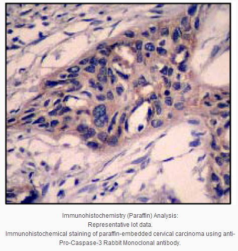
Immunohistochemistry Analysis:
1:25 - 50 dilution from a previous lot detected Caspase-3 in human cervical carcinoma tissue.
Biological Information
Immunogen
Synthetic peptide corresponding to residues in p17 subunit of human Caspase-3.
Epitope
p17 subunit
Clone
E87
Host
Rabbit
Specificity
The antibody should recognize both pro-form (35 kDa) and p17 cleaved-form of Caspase-3.
This gene encodes a protein which is a member of the cysteine-aspartic acid protease (caspase) family. Sequential activation of caspases plays a central role in the execution-phase of cell apoptosis. Caspases exist as inactive proenzymes which undergo proteolytic processing at conserved aspartic residues to produce two subunits, large and small, that dimerize to form the active enzyme. This protein cleaves and activates caspases 6, 7 and 9, and the protein itself is processed by caspases 8, 9 and 10. It is the predominant caspase involved in the cleavage of amyloid-beta 4A precursor protein, which is associated with neuronal death in Alzheimer's disease. Alternative splicing of this gene results in two transcript variants that encode the same protein. [provided by RefSeq].
FUNCTION:Involved in the activation cascade of caspases responsible for apoptosis execution. At the onset of apoptosis it proteolytically cleaves poly(ADP-ribose) polymerase (PARP) at a '216-Asp-|-Gly-217' bond. Cleaves and activates sterol regulatory element binding proteins (SREBPs) between the basic helix-loop-helix leucine zipper domain and the membrane attachment domain. Cleaves and activates caspase-6, -7 and -9. Involved in the cleavage of huntingtin.
CATALYTIC ACTIVITY: Strict requirement for an Asp residue at positions P1 and P4. It has a preferred cleavage sequence of Asp-Xaa-Xaa-Asp-|- with a hydrophobic amino-acid residue at P2 and a hydrophilic amino-acid residue at P3, although Val or Ala are also accepted at this position.
ENZYME REGULATION:Inhibited by isatin sulfonamides.
SUBUNIT STRUCTURE:Heterotetramer that consists of two anti-parallel arranged heterodimers, each one formed by a 17 kDa (p17) and a 12 kDa (p12) subunit.
Subcellular location Cytoplasm.
TISSUE SPECIFICITY:Highly expressed in lung, spleen, heart, liver and kidney. Moderate levels in brain and skeletal muscle, and low in testis. Also found in many cell lines, highest expression in cells of the immune system.
PTM:Cleavage by granzyme B, caspase-6, caspase-8 and caspase-10 generates the two active subunits. Additional processing of the propeptides is likely due to the autocatalytic activity of the activated protease. Active heterodimers between the small subunit of caspase-7 protease and the large subunit of caspase-3 also occur and vice versa.
S-nitrosylated on its catalytic site cysteine in unstimulated human cell lines and denitrosylated upon activation of the Fas apoptotic pathway, associated with an increase in intracellular caspase activity. Fas therefore activates caspase-3 not only by inducing the cleavage of the caspase zymogen to its active subunits, but also by stimulating the denitrosylation of its active site thiol.
SEQUENCE SIMILARITY:Belongs to the peptidase C14A family.
Product Usage Statements
Quality Assurance
Evaluated by Western Blotting on Jurkat cell lysates.
Western Blotting Analysis:
1:5,000 dilution of this antibody was used to detect Caspase-3 in Jurkat cell lysate.
Usage Statement
Unless otherwise stated in our catalog or other company documentation accompanying the product(s), our products are intended for research use only and are not to be used for any other purpose, which includes but is not limited to, unauthorized commercial uses, in vitro diagnostic uses, ex vivo or in vivo therapeutic uses or any type of consumption or application to humans or animals.
Storage and Shipping Information
Storage Conditions
Stable for 1 year at -20ºC from date of receipt.
Handling Recommendations: Upon receipt, and prior to removing the cap, centrifuge the vial and gently mix the solution. Aliquot into microcentrifuge tubes and store at -20°C. Avoid repeated freeze/thaw cycles, which may damage IgG and affect product performance. Note: Variability in freezer temperatures below -20°C may cause glycerol containing solutions to become frozen during storage.
Caspase-3 (CPP32, Apopain) is the most extensively studied apoptotic protein. Caspase-3 is synthesized as an inactive proenzyme (32 kDa) that is processed in cells undergoing apoptosis by self-proteolysis and/or cleavage by another upstream protease. The processed form of caspase-3 consists of large (17 kDa) and small (12 kDa) subunits which associate to form an active enzyme. The active caspase-3 proteolytically cleaves and activates other caspases, as well as relevant targets in the cells such as PARP and DFF.
Product Information
Format
Unpurified
Control
Jurkat lysates
Presentation
Rabbit Monoclonal in buffer containing 50 mM Tris-Glycine (pH 7.4), 0.15 M NaCl containing 40% Glycerol, 0.01% sodium azide and 0.05% BSA.
Applications
Application
Please note that this product will not be available for sale after March 15, 2015. Please select one of the other antibodies against this target. Anti-Caspase-3 Antibody, clone E87, Rabbit is an antibody against Caspase-3 for use in WB, IP, IH(P) & IC.
Key Applications
Immunocytochemistry
Western Blotting
Immunoprecipitation
Immunohistochemistry (Paraffin)
Application Notes
Immunohistochemistry Analysis:
1:25 - 50 dilution from a previous lot detected Caspase-3 in human cervical carcinoma tissue.
Biological Information
Immunogen
Synthetic peptide corresponding to residues in p17 subunit of human Caspase-3.
Epitope
p17 subunit
Clone
E87
Host
Rabbit
Specificity
The antibody should recognize both pro-form (35 kDa) and p17 cleaved-form of Caspase-3.
This gene encodes a protein which is a member of the cysteine-aspartic acid protease (caspase) family. Sequential activation of caspases plays a central role in the execution-phase of cell apoptosis. Caspases exist as inactive proenzymes which undergo proteolytic processing at conserved aspartic residues to produce two subunits, large and small, that dimerize to form the active enzyme. This protein cleaves and activates caspases 6, 7 and 9, and the protein itself is processed by caspases 8, 9 and 10. It is the predominant caspase involved in the cleavage of amyloid-beta 4A precursor protein, which is associated with neuronal death in Alzheimer's disease. Alternative splicing of this gene results in two transcript variants that encode the same protein. [provided by RefSeq].
FUNCTION:Involved in the activation cascade of caspases responsible for apoptosis execution. At the onset of apoptosis it proteolytically cleaves poly(ADP-ribose) polymerase (PARP) at a '216-Asp-|-Gly-217' bond. Cleaves and activates sterol regulatory element binding proteins (SREBPs) between the basic helix-loop-helix leucine zipper domain and the membrane attachment domain. Cleaves and activates caspase-6, -7 and -9. Involved in the cleavage of huntingtin.
CATALYTIC ACTIVITY: Strict requirement for an Asp residue at positions P1 and P4. It has a preferred cleavage sequence of Asp-Xaa-Xaa-Asp-|- with a hydrophobic amino-acid residue at P2 and a hydrophilic amino-acid residue at P3, although Val or Ala are also accepted at this position.
ENZYME REGULATION:Inhibited by isatin sulfonamides.
SUBUNIT STRUCTURE:Heterotetramer that consists of two anti-parallel arranged heterodimers, each one formed by a 17 kDa (p17) and a 12 kDa (p12) subunit.
Subcellular location Cytoplasm.
TISSUE SPECIFICITY:Highly expressed in lung, spleen, heart, liver and kidney. Moderate levels in brain and skeletal muscle, and low in testis. Also found in many cell lines, highest expression in cells of the immune system.
PTM:Cleavage by granzyme B, caspase-6, caspase-8 and caspase-10 generates the two active subunits. Additional processing of the propeptides is likely due to the autocatalytic activity of the activated protease. Active heterodimers between the small subunit of caspase-7 protease and the large subunit of caspase-3 also occur and vice versa.
S-nitrosylated on its catalytic site cysteine in unstimulated human cell lines and denitrosylated upon activation of the Fas apoptotic pathway, associated with an increase in intracellular caspase activity. Fas therefore activates caspase-3 not only by inducing the cleavage of the caspase zymogen to its active subunits, but also by stimulating the denitrosylation of its active site thiol.
SEQUENCE SIMILARITY:Belongs to the peptidase C14A family.
Product Usage Statements
Quality Assurance
Evaluated by Western Blotting on Jurkat cell lysates.
Western Blotting Analysis:
1:5,000 dilution of this antibody was used to detect Caspase-3 in Jurkat cell lysate.
Usage Statement
Unless otherwise stated in our catalog or other company documentation accompanying the product(s), our products are intended for research use only and are not to be used for any other purpose, which includes but is not limited to, unauthorized commercial uses, in vitro diagnostic uses, ex vivo or in vivo therapeutic uses or any type of consumption or application to humans or animals.
Storage and Shipping Information
Storage Conditions
Stable for 1 year at -20ºC from date of receipt.
Handling Recommendations: Upon receipt, and prior to removing the cap, centrifuge the vial and gently mix the solution. Aliquot into microcentrifuge tubes and store at -20°C. Avoid repeated freeze/thaw cycles, which may damage IgG and affect product performance. Note: Variability in freezer temperatures below -20°C may cause glycerol containing solutions to become frozen during storage.


 京公网安备11010802025653 版权所有:北京逸优科技有限公司
