Members of the SMAD family of signal transduction molecules are components of a critical intracellular pathway that transmits TGF-β signals from the cell surface into the nucleus. Three distinct classes of SMADs have been defined: the receptor-regulated SMADs (R-SMADs), which include Smad1, 2, 3, 5 and 8, the common-mediator SMAD (co-SMAD), SMAD4, and the antagonistic or inhibitory SMADs (I-SMADs), SMAD6 and 7. Once in the nucleus, SMADs can target a variety of DNA binding proteins to regulate transcriptional responses. Following stimulation by TGF-β, SMAD2 and SMAD3 become phosphorylated at their carboxyl termini (Ser465 and 467 on SMAD2; Ser423 and 425 on SMAD3) by TGF-β Receptor I. Phosphorylated SMAD 2/3 can complex with SMAD4, translocate to the nucleus and regulate gene expression.
Product Information
Format
Unpurified
Control
Jurkat cell lysate
Presentation
Rabbit Monoclonal in buffer containing 50 mM Tris-Glycine (pH 7.4), 0.15 M NaCl containing 40% Glycerol, 0.01% sodium azide and 0.05% BSA.
Applications
Application
Please note that this product will not be available for sale after March 15, 2015. Please select one of the other antibodies against this target. Anti-SMAD3 Antibody, clone EP568Y, Rabbit is an antibody against SMAD3 for use in WB, IH(P), IC & IP.
Key Applications
Immunoprecipitation
Western Blotting
Immunohistochemistry (Paraffin)
Immunocytochemistry
Application Notes
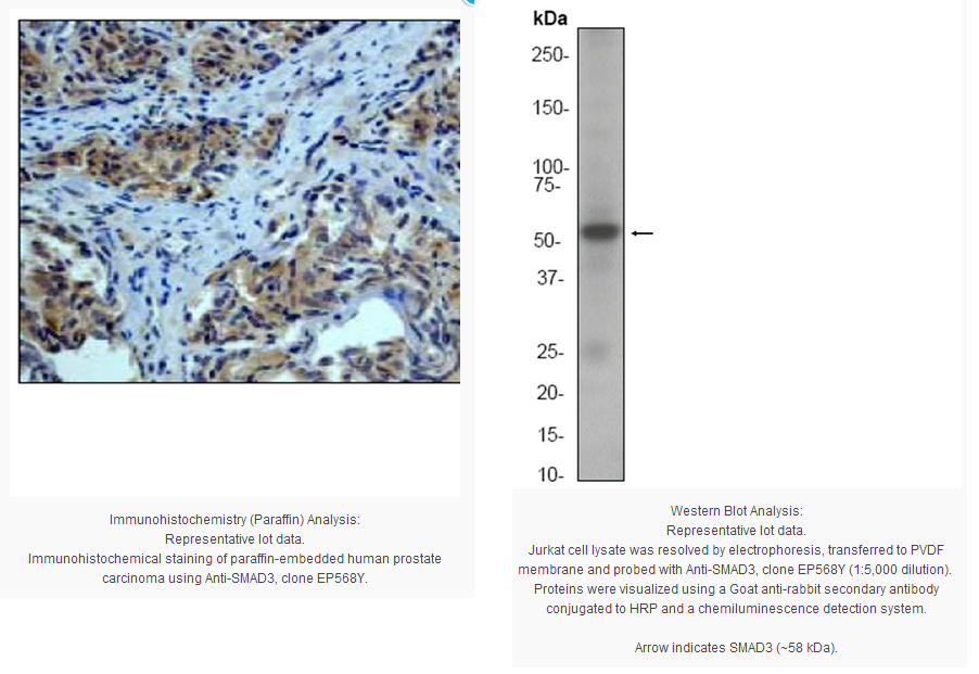
Immunohistochemistry Analysis (paraffin): A 1:100-250 dilution from a previous lot detected SMAD3 in human prostate carcinoma tissue.
Immunocytochemistry Analysis: A 1:100-250 dilution from a previous lot worked in IC.
Immunoprecipitation: A 1:50 dilution from a previous lot was used in IP.
Biological Information
Immunogen
Synthetic peptide corresponding to an internal region of human SMAD3.
FUNCTION:Transcriptional modulator activated by TGF-beta (transforming growth factor) and activin type 1 receptor kinase. SMAD3 is a receptor-regulated SMAD (R-SMAD) By similarity. SUBUNIT STRUCTURE:Interacts with HGS. Interacts with NEDD4L in response to TGF-beta. Interacts with TTRAP By similarity. Interacts with SARA (SMAD anchor for receptor activation); form trimers with another SMAD3 and the co-SMAD SMAD4. Interacts with JUN/FOS, vitamin D receptor, homeobox protein TGIF and TGIF2, PEBP2-alpha C subunit, CREB-binding protein (CBP), p300, SKI, SNON, ATF2, SMURF2, AIP1, DACH1 and TGFB1I1. Part of a complex consisting of AIP1, ACVR2A, ACVR1B and SMAD3. Found in a complex with SMAD2 and TRIM33 upon addition of TGF-beta. Interacts with SMAD2 and TRIM33. Found in a complex with SMAD3, Ran and XPO4. Interacts with XPO4. Interacts with LBXCOR1 and CORL2. SUBCELLULAR LOCATION:Cytoplasm By similarity. Nucleus By similarity. Note: In the cytoplasm in the absence of ligand. Migration to the nucleus when complexed with SMAD4 By similarity. DOMAIN:The MH2 domain is sufficient to carry protein nuclear export. PTM:Phosphorylated on serine by TGF-beta and activin type 1 receptor kinases. INVOLVEMENT IN DISEASE:Defects in SMAD3 may be a cause of colorectal cancer (CRC) [MIM:114500]. SEQUENCE SIMILARITIES:Belongs to the dwarfin/SMAD family.
Contains 1 MH1 (MAD homology 1) domain.
Contains 1 MH2 (MAD homology 2) domain.
Product Usage Statements
Quality Assurance
Evaluated by Western Blot on Jurkat cell lysates.
Western Blot Analysis: A 1:5,000 dilution of this antibody was used to detect SMAD3 in Jurkat cell lysate.
Usage Statement
Unless otherwise stated in our catalog or other company documentation accompanying the product(s), our products are intended for research use only and are not to be used for any other purpose, which includes but is not limited to, unauthorized commercial uses, in vitro diagnostic uses, ex vivo or in vivo therapeutic uses or any type of consumption or application to humans or animals.
Storage and Shipping Information
Storage Conditions
Stable for 1 year at -20ºC from date of receipt.
Handling Recommendations: Upon receipt and prior to removing the cap, centrifuge the vial and gently mix the solution. Aliquot into microcentrifuge tubes and store at -20°C. Avoid repeated freeze/thaw cycles, which may damage IgG and affect product performance.
Note: Variability in freezer temperatures below -20°C may cause glycerol containing solutions to become frozen during storage.
Members of the SMAD family of signal transduction molecules are components of a critical intracellular pathway that transmits TGF-β signals from the cell surface into the nucleus. Three distinct classes of SMADs have been defined: the receptor-regulated SMADs (R-SMADs), which include Smad1, 2, 3, 5 and 8, the common-mediator SMAD (co-SMAD), SMAD4, and the antagonistic or inhibitory SMADs (I-SMADs), SMAD6 and 7. Once in the nucleus, SMADs can target a variety of DNA binding proteins to regulate transcriptional responses. Following stimulation by TGF-β, SMAD2 and SMAD3 become phosphorylated at their carboxyl termini (Ser465 and 467 on SMAD2; Ser423 and 425 on SMAD3) by TGF-β Receptor I. Phosphorylated SMAD 2/3 can complex with SMAD4, translocate to the nucleus and regulate gene expression.
Product Information
Format
Unpurified
Control
Jurkat cell lysate
Presentation
Rabbit Monoclonal in buffer containing 50 mM Tris-Glycine (pH 7.4), 0.15 M NaCl containing 40% Glycerol, 0.01% sodium azide and 0.05% BSA.
Applications
Application
Please note that this product will not be available for sale after March 15, 2015. Please select one of the other antibodies against this target. Anti-SMAD3 Antibody, clone EP568Y, Rabbit is an antibody against SMAD3 for use in WB, IH(P), IC & IP.
Key Applications
Immunoprecipitation
Western Blotting
Immunohistochemistry (Paraffin)
Immunocytochemistry
Application Notes
Immunohistochemistry Analysis (paraffin): A 1:100-250 dilution from a previous lot detected SMAD3 in human prostate carcinoma tissue.
Immunocytochemistry Analysis: A 1:100-250 dilution from a previous lot worked in IC.
Immunoprecipitation: A 1:50 dilution from a previous lot was used in IP.
Biological Information
Immunogen
Synthetic peptide corresponding to an internal region of human SMAD3.
FUNCTION:Transcriptional modulator activated by TGF-beta (transforming growth factor) and activin type 1 receptor kinase. SMAD3 is a receptor-regulated SMAD (R-SMAD) By similarity. SUBUNIT STRUCTURE:Interacts with HGS. Interacts with NEDD4L in response to TGF-beta. Interacts with TTRAP By similarity. Interacts with SARA (SMAD anchor for receptor activation); form trimers with another SMAD3 and the co-SMAD SMAD4. Interacts with JUN/FOS, vitamin D receptor, homeobox protein TGIF and TGIF2, PEBP2-alpha C subunit, CREB-binding protein (CBP), p300, SKI, SNON, ATF2, SMURF2, AIP1, DACH1 and TGFB1I1. Part of a complex consisting of AIP1, ACVR2A, ACVR1B and SMAD3. Found in a complex with SMAD2 and TRIM33 upon addition of TGF-beta. Interacts with SMAD2 and TRIM33. Found in a complex with SMAD3, Ran and XPO4. Interacts with XPO4. Interacts with LBXCOR1 and CORL2. SUBCELLULAR LOCATION:Cytoplasm By similarity. Nucleus By similarity. Note: In the cytoplasm in the absence of ligand. Migration to the nucleus when complexed with SMAD4 By similarity. DOMAIN:The MH2 domain is sufficient to carry protein nuclear export. PTM:Phosphorylated on serine by TGF-beta and activin type 1 receptor kinases. INVOLVEMENT IN DISEASE:Defects in SMAD3 may be a cause of colorectal cancer (CRC) [MIM:114500]. SEQUENCE SIMILARITIES:Belongs to the dwarfin/SMAD family.
Contains 1 MH1 (MAD homology 1) domain.
Contains 1 MH2 (MAD homology 2) domain.
Product Usage Statements
Quality Assurance
Evaluated by Western Blot on Jurkat cell lysates.
Western Blot Analysis: A 1:5,000 dilution of this antibody was used to detect SMAD3 in Jurkat cell lysate.
Usage Statement
Unless otherwise stated in our catalog or other company documentation accompanying the product(s), our products are intended for research use only and are not to be used for any other purpose, which includes but is not limited to, unauthorized commercial uses, in vitro diagnostic uses, ex vivo or in vivo therapeutic uses or any type of consumption or application to humans or animals.
Storage and Shipping Information
Storage Conditions
Stable for 1 year at -20ºC from date of receipt.
Handling Recommendations: Upon receipt and prior to removing the cap, centrifuge the vial and gently mix the solution. Aliquot into microcentrifuge tubes and store at -20°C. Avoid repeated freeze/thaw cycles, which may damage IgG and affect product performance.
Note: Variability in freezer temperatures below -20°C may cause glycerol containing solutions to become frozen during storage.


 京公网安备11010802025653 版权所有:北京逸优科技有限公司
