Neurofilament-66, also known as α-internexin, is a member of the IF family. It is found in developing neuroblasts and in the CNS of adults. Neurofilament-66 is expressed in the adult brain at lower levels than other neurofilament proteins.
Product Information
Format
Unpurified
Control
SHSY5Y cell lysate
Presentation
Rabbit Monoclonal in buffer containing 50 mM Tris-Glycine (pH 7.4), 0.15 M NaCl containing 40% Glycerol, 0.01% sodium azide and 0.05% BSA.
Applications
Application
Please note that this product will not be available for sale after March 15, 2015. Please select one of the other antibodies against this target.
Key Applications
Flow Cytometry
Western Blotting
Immunohistochemistry (Paraffin)
Immunocytochemistry
Immunoprecipitation
Application Notes
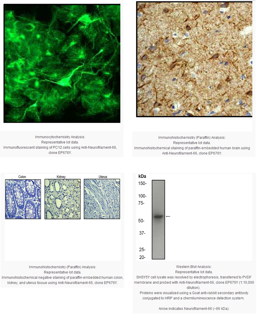
Immunohistochemistry Analysis: A 1:250-500 dilution from a previous lot detected Neurofilament-66 in human brain tissue but not in human colon, kidney or uterus.
Immunocytochemistry Analysis: A 1:250 - 500 dilution from a previous lot detected Neurofilament-66 in PC12 cells.
Flow Cytometry: A 1:25 dilution of a previous lot was used in flow cytometry.
Immunoprecipitation Analysis: A 1:50 dilution from a previous lot was used in IP.
Biological Information
Immunogen
Synthetic peptide corresponding to residues in the tail domain of human Neurofilament-66.
Epitope
Tail Domain
Clone
EP676Y
Host
Rabbit
Specificity
This antibody recognizes Neurofilament-66 at and around the tail domain.
Neurofilaments are type IV intermediate filament heteropolymers composed of light, medium, and heavy chains. Neurofilaments comprise the axoskeleton and they functionally maintain the neuronal caliber. They may also play a role in intracellular transport to axons and dendrites. This gene is a member of the intermediate filament family and is involved in the morphogenesis of neurons.
FUNCTION:Class-IV neuronal intermediate filament that is able to self-assemble. It is involved in the morphogenesis of neurons. It may form an independent structural network without the involvement of other neurofilaments or it may cooperate with NF-L to form the filamentous backbone to which NF-M and NF-H attach to form the cross-bridges. TISSUE SPECIFICITY:Found predominantly in adult CNS. DEVELOPMENTAL STAGE:Expressed in brain as early as the 16th week of gestation, and increased rapidly and reached a steady state level by the 18th week of gestation. PTM:O-glycosylated By similarity.
Phosphorylated upon DNA damage, probably by ATM or ATR. SEQUENCE SIMILARITIES:Belongs to the intermediate filament family.
Product Usage Statements
Quality Assurance
Evaluated by Western Blot on SHSY5Y cell lysates.
Western Blot Analysis: 1:5,000-10,000 dilution of this antibody was used to detect Neurofilament-66 in SHSY5Y cell lysate.
Usage Statement
Unless otherwise stated in our catalog or other company documentation accompanying the product(s), our products are intended for research use only and are not to be used for any other purpose, which includes but is not limited to, unauthorized commercial uses, in vitro diagnostic uses, ex vivo or in vivo therapeutic uses or any type of consumption or application to humans or animals.
Storage and Shipping Information
Storage Conditions
Stable for 1 year at -20ºC from date of receipt.
Handling Recommendations: Upon first thaw, and prior to removing the cap, centrifuge the vial and gently mix the solution. Aliquot into microcentrifuge tubes and store at -20°C. Avoid repeated freeze/thaw cycles, which may damage IgG and affect product performance. Note: Variability in freezer temperatures below -20°C may cause glycerol containing solutions to become frozen during storage.
Neurofilament-66, also known as α-internexin, is a member of the IF family. It is found in developing neuroblasts and in the CNS of adults. Neurofilament-66 is expressed in the adult brain at lower levels than other neurofilament proteins.
Product Information
Format
Unpurified
Control
SHSY5Y cell lysate
Presentation
Rabbit Monoclonal in buffer containing 50 mM Tris-Glycine (pH 7.4), 0.15 M NaCl containing 40% Glycerol, 0.01% sodium azide and 0.05% BSA.
Applications
Application
Please note that this product will not be available for sale after March 15, 2015. Please select one of the other antibodies against this target.
Key Applications
Flow Cytometry
Western Blotting
Immunohistochemistry (Paraffin)
Immunocytochemistry
Immunoprecipitation
Application Notes
Immunohistochemistry Analysis: A 1:250-500 dilution from a previous lot detected Neurofilament-66 in human brain tissue but not in human colon, kidney or uterus.
Immunocytochemistry Analysis: A 1:250 - 500 dilution from a previous lot detected Neurofilament-66 in PC12 cells.
Flow Cytometry: A 1:25 dilution of a previous lot was used in flow cytometry.
Immunoprecipitation Analysis: A 1:50 dilution from a previous lot was used in IP.
Biological Information
Immunogen
Synthetic peptide corresponding to residues in the tail domain of human Neurofilament-66.
Epitope
Tail Domain
Clone
EP676Y
Host
Rabbit
Specificity
This antibody recognizes Neurofilament-66 at and around the tail domain.
Neurofilaments are type IV intermediate filament heteropolymers composed of light, medium, and heavy chains. Neurofilaments comprise the axoskeleton and they functionally maintain the neuronal caliber. They may also play a role in intracellular transport to axons and dendrites. This gene is a member of the intermediate filament family and is involved in the morphogenesis of neurons.
FUNCTION:Class-IV neuronal intermediate filament that is able to self-assemble. It is involved in the morphogenesis of neurons. It may form an independent structural network without the involvement of other neurofilaments or it may cooperate with NF-L to form the filamentous backbone to which NF-M and NF-H attach to form the cross-bridges. TISSUE SPECIFICITY:Found predominantly in adult CNS. DEVELOPMENTAL STAGE:Expressed in brain as early as the 16th week of gestation, and increased rapidly and reached a steady state level by the 18th week of gestation. PTM:O-glycosylated By similarity.
Phosphorylated upon DNA damage, probably by ATM or ATR. SEQUENCE SIMILARITIES:Belongs to the intermediate filament family.
Product Usage Statements
Quality Assurance
Evaluated by Western Blot on SHSY5Y cell lysates.
Western Blot Analysis: 1:5,000-10,000 dilution of this antibody was used to detect Neurofilament-66 in SHSY5Y cell lysate.
Usage Statement
Unless otherwise stated in our catalog or other company documentation accompanying the product(s), our products are intended for research use only and are not to be used for any other purpose, which includes but is not limited to, unauthorized commercial uses, in vitro diagnostic uses, ex vivo or in vivo therapeutic uses or any type of consumption or application to humans or animals.
Storage and Shipping Information
Storage Conditions
Stable for 1 year at -20ºC from date of receipt.
Handling Recommendations: Upon first thaw, and prior to removing the cap, centrifuge the vial and gently mix the solution. Aliquot into microcentrifuge tubes and store at -20°C. Avoid repeated freeze/thaw cycles, which may damage IgG and affect product performance. Note: Variability in freezer temperatures below -20°C may cause glycerol containing solutions to become frozen during storage.


 京公网安备11010802025653 版权所有:北京逸优科技有限公司
