Synaptophysin is a protein of the presynaptic vesicle exocytosis machinery interacting at the vesicle with VAMP/synaptobrevin. Synaptophysin structure consists of a hexameric protein of four transmembrane spans with cytoplasmic amino and carboxyl termini. Although its actual function remains elusive, synaptophysin remains a specific presynaptic marker for all neurons, neuroendocrine cells and neuroderived neoplasms.
Product Information
Format
Unpurified
Control
PC12 cell lysate
Presentation
Rabbit Monoclonal in buffer containing 50 mM Tris-Glycine (pH 7.4), 0.15 M NaCl containing 40% Glycerol, 0.01% sodium azide and 0.05% BSA.
Applications
Application
Please note that this product will not be available for sale after March 15, 2015. Please select one of the other antibodies against this target. This Anti-Synaptophysin (C-term) Antibody, clone YE269, Rabbit is validated for use in WB, IC, IH for the detection of Synaptophysin (C-term).
Key Applications
Immunohistochemistry
Western Blotting
Immunocytochemistry
Application Notes
Immunohistochemistry: A 1:250-1:500 dilution of a previous lot of this antibody was used in immunohistochemistry.
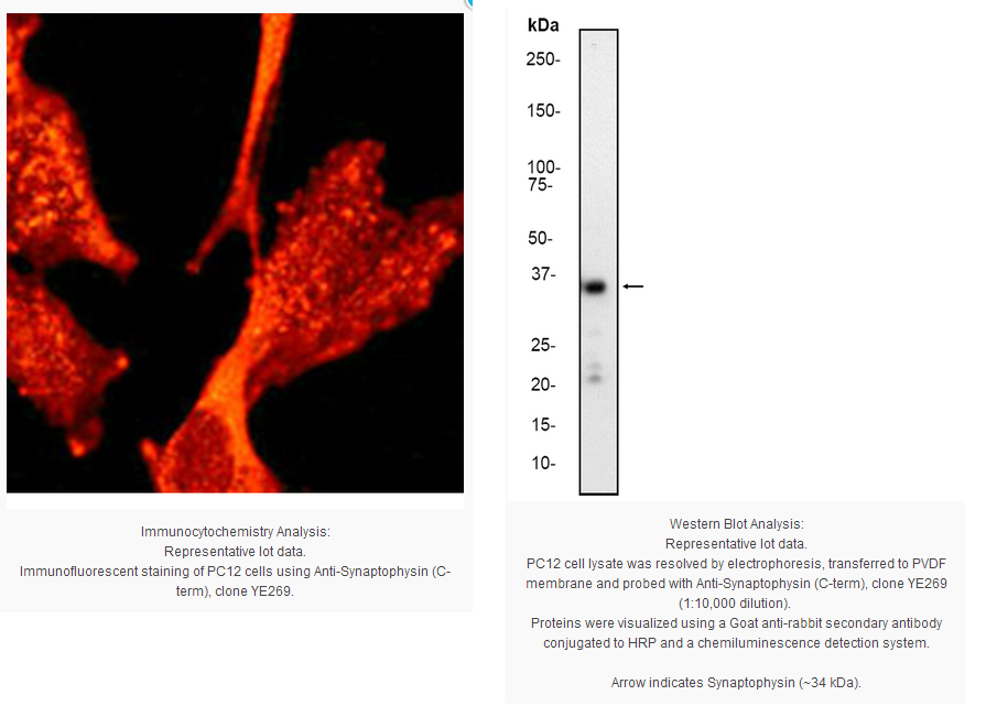
Immunocytochemistry Analysis: A 1:250 dilution of a previous lot was used in immunocytochemistry.
Biological Information
Immunogen
Synthetic peptide corresponding to residues in C-terminus (cytoplasmic domain) of human Synaptophysin.
Epitope
C-terminus
Clone
YE269
Host
Rabbit
Specificity
This antibody recognizes Synaptophysin at and around the c-terminus.
Isotype
IgG
Species Reactivity
Bovine Mouse Rat Human
Species Reactivity Note
Human, mouse and rat. Predicted to cross-react with bovine, based on sequence homology.
FUNCTION:Possibly involved in structural functions as organizing other membrane components or in targeting the vesicles to the plasma membrane. SUBUNIT STRUCTURE:Homohexamer or homotetramer. SUBCELLULAR LOCATION:Cytoplasmic vesicle › secretory vesicle › synaptic vesicle membrane; Multi-pass membrane protein. Cell junction › synapse › synaptosome. TISSUE SPECIFICITY:Characteristic of a type of small (30-80 nm) neurosecretory vesicles, including presynaptic vesicles, but also vesicles of various neuroendocrine cells of both neuronal and epithelial phenotype. DOMAIN:The calcium-binding activity is thought to be localized in the cytoplasmic tail of the protein. PTM:Ubiquitinated; mediated by SIAH1 or SIAH2 and leading to its subsequent proteasomal degradation By similarity. SEQUENCE SIMILARIITES:Belongs to the synaptophysin/synaptobrevin family.
Contains 1 MARVEL domain.
Product Usage Statements
Quality Assurance
Evaluated by Western Blot on PC12 cell lysates.
Western Blot Analysis: 1:10,000 dilution of this antibody was used to detect Synaptophysin in PC12 cell lysate.
Usage Statement
Unless otherwise stated in our catalog or other company documentation accompanying the product(s), our products are intended for research use only and are not to be used for any other purpose, which includes but is not limited to, unauthorized commercial uses, in vitro diagnostic uses, ex vivo or in vivo therapeutic uses or any type of consumption or application to humans or animals.
Storage and Shipping Information
Storage Conditions
Stable for 1 year at -20ºC from date of receipt.
Handling Recommendations: Upon first thaw, and prior to removing the cap, centrifuge the vial and gently mix the solution. Aliquot into microcentrifuge tubes and store at -20°C. Avoid repeated freeze/thaw cycles, which may damage IgG and affect product performance. Note: Variability in freezer temperatures below -20°C may cause glycerol containing solutions to become frozen during storage.
Synaptophysin is a protein of the presynaptic vesicle exocytosis machinery interacting at the vesicle with VAMP/synaptobrevin. Synaptophysin structure consists of a hexameric protein of four transmembrane spans with cytoplasmic amino and carboxyl termini. Although its actual function remains elusive, synaptophysin remains a specific presynaptic marker for all neurons, neuroendocrine cells and neuroderived neoplasms.
Product Information
Format
Unpurified
Control
PC12 cell lysate
Presentation
Rabbit Monoclonal in buffer containing 50 mM Tris-Glycine (pH 7.4), 0.15 M NaCl containing 40% Glycerol, 0.01% sodium azide and 0.05% BSA.
Applications
Application
Please note that this product will not be available for sale after March 15, 2015. Please select one of the other antibodies against this target. This Anti-Synaptophysin (C-term) Antibody, clone YE269, Rabbit is validated for use in WB, IC, IH for the detection of Synaptophysin (C-term).
Key Applications
Immunohistochemistry
Western Blotting
Immunocytochemistry
Application Notes
Immunohistochemistry: A 1:250-1:500 dilution of a previous lot of this antibody was used in immunohistochemistry.
Immunocytochemistry Analysis: A 1:250 dilution of a previous lot was used in immunocytochemistry.
Biological Information
Immunogen
Synthetic peptide corresponding to residues in C-terminus (cytoplasmic domain) of human Synaptophysin.
Epitope
C-terminus
Clone
YE269
Host
Rabbit
Specificity
This antibody recognizes Synaptophysin at and around the c-terminus.
Isotype
IgG
Species Reactivity
Bovine Mouse Rat Human
Species Reactivity Note
Human, mouse and rat. Predicted to cross-react with bovine, based on sequence homology.
FUNCTION:Possibly involved in structural functions as organizing other membrane components or in targeting the vesicles to the plasma membrane. SUBUNIT STRUCTURE:Homohexamer or homotetramer. SUBCELLULAR LOCATION:Cytoplasmic vesicle › secretory vesicle › synaptic vesicle membrane; Multi-pass membrane protein. Cell junction › synapse › synaptosome. TISSUE SPECIFICITY:Characteristic of a type of small (30-80 nm) neurosecretory vesicles, including presynaptic vesicles, but also vesicles of various neuroendocrine cells of both neuronal and epithelial phenotype. DOMAIN:The calcium-binding activity is thought to be localized in the cytoplasmic tail of the protein. PTM:Ubiquitinated; mediated by SIAH1 or SIAH2 and leading to its subsequent proteasomal degradation By similarity. SEQUENCE SIMILARIITES:Belongs to the synaptophysin/synaptobrevin family.
Contains 1 MARVEL domain.
Product Usage Statements
Quality Assurance
Evaluated by Western Blot on PC12 cell lysates.
Western Blot Analysis: 1:10,000 dilution of this antibody was used to detect Synaptophysin in PC12 cell lysate.
Usage Statement
Unless otherwise stated in our catalog or other company documentation accompanying the product(s), our products are intended for research use only and are not to be used for any other purpose, which includes but is not limited to, unauthorized commercial uses, in vitro diagnostic uses, ex vivo or in vivo therapeutic uses or any type of consumption or application to humans or animals.
Storage and Shipping Information
Storage Conditions
Stable for 1 year at -20ºC from date of receipt.
Handling Recommendations: Upon first thaw, and prior to removing the cap, centrifuge the vial and gently mix the solution. Aliquot into microcentrifuge tubes and store at -20°C. Avoid repeated freeze/thaw cycles, which may damage IgG and affect product performance. Note: Variability in freezer temperatures below -20°C may cause glycerol containing solutions to become frozen during storage.


 京公网安备11010802025653 版权所有:北京逸优科技有限公司
