Hypoxia, a condition of low tissue O2 concentration, plays an important role in normal physiological processes and tumor formation. Under hypoxic conditions mammalian cells up regulate the expression of hypoxic genes, including induction of angiogenesis and a switch to anaerobic metabolism, in order to survive. HIF-1 (Hypoxia Inducible Factor-1) is one of the key regulators of the transcriptional response to oxygen deprivation (1). HIF-1 is composed of two subunits, HIF-1αand HIF-1β also known as aryl hydrocarbon receptor nuclear translocator (ARNT)) that are members of the basic helix-loop-helix (bHLH) Per-Arnt-Sim (PAS) (bHLH-PAS) family of transcription factors. HIF-1 is essential for angiogenesis, embryonic development, and is associated with tumor progression, erythropoiesis, vascular development/remodeling, vasodilation, and glucose/energy metabolism. The over expression of HIF-1αhas been demonstrated in many common human cancers including prostate and breast, in which HIF-1α levels are associated with increase vascularitry and tumor progression. Besides physiological hypoxia, genetic abnormalities frequently detected in human cancers, such as loss of function mutations (Von Hippel-Lindau, p53, and PTEN), are associated with induction of HIF1 activity and expression of HIF-1-inducible genes (1).
Product Information
Format
Unpurified
Control
Ramos + CoCl2 lysate
Presentation
Rabbit Monoclonal in buffer containing 50 mM Tris-Glycine (pH 7.4), 0.15 M NaCl containing 40% Glycerol, 0.01% sodium azide and 0.05% BSA.
Applications
Application
Please note that this product will not be available for sale after March 15, 2015. Please select one of the other antibodies against this target.
Key Applications
Immunohistochemistry (Paraffin)
Western Blotting
Immunocytochemistry
Application Notes
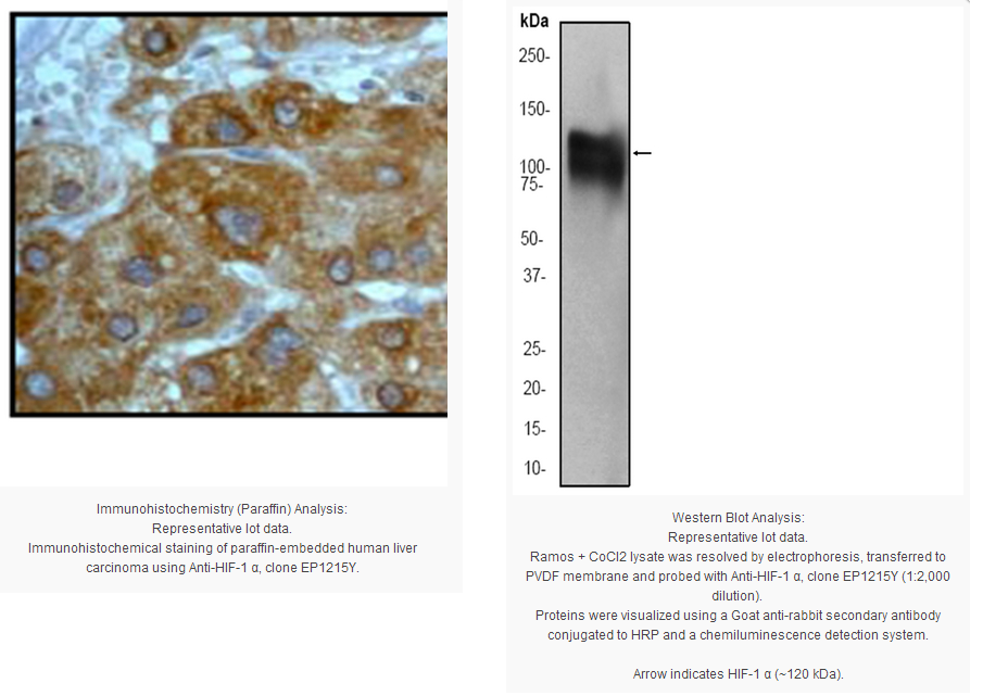
Immunohistochemistry Analysis: A 1:100-250 dilution from a previous lot detected HIF-1α in human liver carcinoma tissue.
Immunocytochemistry Analysis: A 1:100-250 dilution from a previous lot was used in IC.
Biological Information
Immunogen
Synthetic peptide corresponding to residues near the C-terminus of human HIF-1α.
Epitope
C-terminus
Clone
EP1215Y
Host
Rabbit
Specificity
This antibody recognizes HIF-1α at and around the C-terminus.
FUNCTION:Functions as a master transcriptional regulator of the adaptive response to hypoxia. Under hypoxic conditions activates the transcription of over 40 genes, including, erythropoietin, glucose transporters, glycolytic enzymes, vascular endothelial growth factor, and other genes whose protein products increase oxygen delivery or facilitate metabolic adaptation to hypoxia. Plays an essential role in embryonic vascularization, tumor angiogenesis and pathophysiology of ischemic disease. Binds to core DNA sequence 5'-[AG]CGTG-3' within the hypoxia response element (HRE) of target gene promoters. Activation requires recruitment of transcriptional coactivators such as CREBPB and EP300. Activity is enhanced by interaction with both, NCOA1 or NCOA2. Interaction with redox regulatory protein APEX seems to activate CTAD and potentiates activation by NCOA1 and CREBBP. SUBUNIT STRUCTURE:Interacts with the HIF1A beta/ARNT subunit; heterodimerization is required for DNA binding. Interacts with COPS5; the interaction increases the transcriptional activity of HIF1A through increased stability By similarity. Interacts with CREBBP and EP300 (via TAZ-type 1 domains). Interacts with NCOA1, NCOA2, APEX and HSP90. Interacts (hydroxylated within the ODD domain) with VHLL (via beta domain); the interaction, leads to polyubiquitination and subsequent HIF1A proteasomal degradation. During hypoxia, sumoylated HIF1A also binds VHL; the interaction promotes the ubiquitination of HIF1A. Interacts with SENP1; the interaction desumoylates HIF1A resulting in stabilization and activation of transcription. Interacts (Via the ODD domain) with ARD1A; the interaction appears not to acetylate HIF1A nor have any affect on protein stability, during hypoxia. Interacts with RWDD3; the interaction enhances HIF1A sumoylation. Interacts with TSGA10 By similarity. SUBCELLULAR LOCATION:Cytoplasm. Nucleus. Note: Cytoplasmic in normoxia, nuclear translocation in response to hypoxia. Colocalizes with SUMO1 in the nucleus, under hypoxia. TISSUE SPECIFICITY:Expressed in most tissues with highest levels in kidney and heart. Overexpressed in the majority of common human cancers and their metastases, due to the presence of intratumoral hypoxia and as a result of mutations in genes encoding oncoproteins and tumor suppressors. INDUCTION:Under reduced oxygen tension. Induced also by various receptor-mediated factors such as growth factors, cytokines, and circulatory factors such as PDGF, EGF FGF-2 FGF-2 IGF-2, TGF-1 beta, HGF, TNF alpha, IL-1 beta, angiotensin-2 and thrombin. However, this induction is less intense than that stimulated by hypoxia. DOMAIN:Contains two independent C-terminal transactivation domains, NTAD and CTAD, which function synergistically. Their transcriptional activity is repressed by an intervening inhibitory domain (ID). PTM:In normoxia, is hydroxylated on Pro-402 and Pro-564 in the oxygen-dependent degradation domain (ODD) by EGLN1/PHD1 and EGLN2/PHD2. EGLN3/PHD3 has also been shown to hydroxylate Pro-564. The hydroxylated prolines promote interaction with VHL, initiating rapid ubiquitination and subsequent proteasomal degradation. Under hypoxia, proline hydroxylation is impaired and ubiquitination is attenuated, resulting in stabilization.
In normoxia, is hydroxylated on Asn-803 by HIF1AN, thus abrogating interaction with CREBBP and EP300 and preventing transcriptional activation. This hydroxylation is inhibited by the Cu/Zn-chelator, Clioquinol.
S-nitrosylation of Cys-800 may be responsible for increased recruitment of p300 coactivator necessary for transcriptional activity of HIF-1 complex.
Requires phosphorylation for DNA-binding.
Sumoylated; by SUMO1 under hypoxia. Sumoylation is enhanced through interaction with RWDD3. Desumoylation by SENP1 leads to increased HIF1A stability and transriptional activity By similarity.
Ubiquitinated; in normoxia, following hydroxylation and interaction with VHL. Lys-532 appears to be the principal site of ubiquitination. Clioquinol, the Cu/Zn-chelator, inhibits ubiquitination through preventing hydroxylation at Asn-803.
The iron and 2-oxoglutarate dependent 3-hydroxylation of asparagine is (S) stereospecific within HIF CTAD domains. SEQUENCE SIMILARITIES:Contains 1 basic helix-loop-helix (bHLH) domain.
Contains 1 PAC (PAS-associated C-terminal) domain.
Contains 2 PAS (PER-ARNT-SIM) domains.
Product Usage Statements
Quality Assurance
Evaluated by Western Blot on Ramos + CoCl2 lysates.
Western Blot Analysis: A 1:2,000 dilution of this antibody was used to detect HIF-1α in Ramos + CoCl2 lysate.
Usage Statement
Unless otherwise stated in our catalog or other company documentation accompanying the product(s), our products are intended for research use only and are not to be used for any other purpose, which includes but is not limited to, unauthorized commercial uses, in vitro diagnostic uses, ex vivo or in vivo therapeutic uses or any type of consumption or application to humans or animals.
Storage and Shipping Information
Storage Conditions
Stable for 1 year at -20ºC from date of receipt. Handling Recommendations: Upon first thaw, and prior to removing the cap, centrifuge the vial and gently mix the solution. Aliquot into microcentrifuge tubes and store at -20°C. Avoid repeated freeze/thaw cycles, which may damage IgG and affect product performance. Note: Variability in freezer temperatures below -20°C may cause glycerol containing solutions to become frozen during storage.
Hypoxia, a condition of low tissue O2 concentration, plays an important role in normal physiological processes and tumor formation. Under hypoxic conditions mammalian cells up regulate the expression of hypoxic genes, including induction of angiogenesis and a switch to anaerobic metabolism, in order to survive. HIF-1 (Hypoxia Inducible Factor-1) is one of the key regulators of the transcriptional response to oxygen deprivation (1). HIF-1 is composed of two subunits, HIF-1αand HIF-1β also known as aryl hydrocarbon receptor nuclear translocator (ARNT)) that are members of the basic helix-loop-helix (bHLH) Per-Arnt-Sim (PAS) (bHLH-PAS) family of transcription factors. HIF-1 is essential for angiogenesis, embryonic development, and is associated with tumor progression, erythropoiesis, vascular development/remodeling, vasodilation, and glucose/energy metabolism. The over expression of HIF-1αhas been demonstrated in many common human cancers including prostate and breast, in which HIF-1α levels are associated with increase vascularitry and tumor progression. Besides physiological hypoxia, genetic abnormalities frequently detected in human cancers, such as loss of function mutations (Von Hippel-Lindau, p53, and PTEN), are associated with induction of HIF1 activity and expression of HIF-1-inducible genes (1).
Product Information
Format
Unpurified
Control
Ramos + CoCl2 lysate
Presentation
Rabbit Monoclonal in buffer containing 50 mM Tris-Glycine (pH 7.4), 0.15 M NaCl containing 40% Glycerol, 0.01% sodium azide and 0.05% BSA.
Applications
Application
Please note that this product will not be available for sale after March 15, 2015. Please select one of the other antibodies against this target.
Key Applications
Immunohistochemistry (Paraffin)
Western Blotting
Immunocytochemistry
Application Notes
Immunohistochemistry Analysis: A 1:100-250 dilution from a previous lot detected HIF-1α in human liver carcinoma tissue.
Immunocytochemistry Analysis: A 1:100-250 dilution from a previous lot was used in IC.
Biological Information
Immunogen
Synthetic peptide corresponding to residues near the C-terminus of human HIF-1α.
Epitope
C-terminus
Clone
EP1215Y
Host
Rabbit
Specificity
This antibody recognizes HIF-1α at and around the C-terminus.
FUNCTION:Functions as a master transcriptional regulator of the adaptive response to hypoxia. Under hypoxic conditions activates the transcription of over 40 genes, including, erythropoietin, glucose transporters, glycolytic enzymes, vascular endothelial growth factor, and other genes whose protein products increase oxygen delivery or facilitate metabolic adaptation to hypoxia. Plays an essential role in embryonic vascularization, tumor angiogenesis and pathophysiology of ischemic disease. Binds to core DNA sequence 5'-[AG]CGTG-3' within the hypoxia response element (HRE) of target gene promoters. Activation requires recruitment of transcriptional coactivators such as CREBPB and EP300. Activity is enhanced by interaction with both, NCOA1 or NCOA2. Interaction with redox regulatory protein APEX seems to activate CTAD and potentiates activation by NCOA1 and CREBBP. SUBUNIT STRUCTURE:Interacts with the HIF1A beta/ARNT subunit; heterodimerization is required for DNA binding. Interacts with COPS5; the interaction increases the transcriptional activity of HIF1A through increased stability By similarity. Interacts with CREBBP and EP300 (via TAZ-type 1 domains). Interacts with NCOA1, NCOA2, APEX and HSP90. Interacts (hydroxylated within the ODD domain) with VHLL (via beta domain); the interaction, leads to polyubiquitination and subsequent HIF1A proteasomal degradation. During hypoxia, sumoylated HIF1A also binds VHL; the interaction promotes the ubiquitination of HIF1A. Interacts with SENP1; the interaction desumoylates HIF1A resulting in stabilization and activation of transcription. Interacts (Via the ODD domain) with ARD1A; the interaction appears not to acetylate HIF1A nor have any affect on protein stability, during hypoxia. Interacts with RWDD3; the interaction enhances HIF1A sumoylation. Interacts with TSGA10 By similarity. SUBCELLULAR LOCATION:Cytoplasm. Nucleus. Note: Cytoplasmic in normoxia, nuclear translocation in response to hypoxia. Colocalizes with SUMO1 in the nucleus, under hypoxia. TISSUE SPECIFICITY:Expressed in most tissues with highest levels in kidney and heart. Overexpressed in the majority of common human cancers and their metastases, due to the presence of intratumoral hypoxia and as a result of mutations in genes encoding oncoproteins and tumor suppressors. INDUCTION:Under reduced oxygen tension. Induced also by various receptor-mediated factors such as growth factors, cytokines, and circulatory factors such as PDGF, EGF FGF-2 FGF-2 IGF-2, TGF-1 beta, HGF, TNF alpha, IL-1 beta, angiotensin-2 and thrombin. However, this induction is less intense than that stimulated by hypoxia. DOMAIN:Contains two independent C-terminal transactivation domains, NTAD and CTAD, which function synergistically. Their transcriptional activity is repressed by an intervening inhibitory domain (ID). PTM:In normoxia, is hydroxylated on Pro-402 and Pro-564 in the oxygen-dependent degradation domain (ODD) by EGLN1/PHD1 and EGLN2/PHD2. EGLN3/PHD3 has also been shown to hydroxylate Pro-564. The hydroxylated prolines promote interaction with VHL, initiating rapid ubiquitination and subsequent proteasomal degradation. Under hypoxia, proline hydroxylation is impaired and ubiquitination is attenuated, resulting in stabilization.
In normoxia, is hydroxylated on Asn-803 by HIF1AN, thus abrogating interaction with CREBBP and EP300 and preventing transcriptional activation. This hydroxylation is inhibited by the Cu/Zn-chelator, Clioquinol.
S-nitrosylation of Cys-800 may be responsible for increased recruitment of p300 coactivator necessary for transcriptional activity of HIF-1 complex.
Requires phosphorylation for DNA-binding.
Sumoylated; by SUMO1 under hypoxia. Sumoylation is enhanced through interaction with RWDD3. Desumoylation by SENP1 leads to increased HIF1A stability and transriptional activity By similarity.
Ubiquitinated; in normoxia, following hydroxylation and interaction with VHL. Lys-532 appears to be the principal site of ubiquitination. Clioquinol, the Cu/Zn-chelator, inhibits ubiquitination through preventing hydroxylation at Asn-803.
The iron and 2-oxoglutarate dependent 3-hydroxylation of asparagine is (S) stereospecific within HIF CTAD domains. SEQUENCE SIMILARITIES:Contains 1 basic helix-loop-helix (bHLH) domain.
Contains 1 PAC (PAS-associated C-terminal) domain.
Contains 2 PAS (PER-ARNT-SIM) domains.
Product Usage Statements
Quality Assurance
Evaluated by Western Blot on Ramos + CoCl2 lysates.
Western Blot Analysis: A 1:2,000 dilution of this antibody was used to detect HIF-1α in Ramos + CoCl2 lysate.
Usage Statement
Unless otherwise stated in our catalog or other company documentation accompanying the product(s), our products are intended for research use only and are not to be used for any other purpose, which includes but is not limited to, unauthorized commercial uses, in vitro diagnostic uses, ex vivo or in vivo therapeutic uses or any type of consumption or application to humans or animals.
Storage and Shipping Information
Storage Conditions
Stable for 1 year at -20ºC from date of receipt. Handling Recommendations: Upon first thaw, and prior to removing the cap, centrifuge the vial and gently mix the solution. Aliquot into microcentrifuge tubes and store at -20°C. Avoid repeated freeze/thaw cycles, which may damage IgG and affect product performance. Note: Variability in freezer temperatures below -20°C may cause glycerol containing solutions to become frozen during storage.


 京公网安备11010802025653 版权所有:北京逸优科技有限公司
