PathScan? Inflammation Multi-Target Sandwich ELISA Kit
 |
|
| 包装: |
|
| 运保温度: |
|
| 到货周期: |
登录后查看 |
| 标准价: |
¥客户可见 |
| 会员价: |
¥客户可见 |
| 积分: |
客户可见 分 |
| |
|
运费与支付说明:
1.含干冰类产品有运费;
2. 必须现金付款或有信用额度的会员才可以直接发货,否则需要等待现金付款信息。 |
|
其它组分:
ELISA Sample Diluent【包装:25 ml,,运保温度:4°C】
STOP Solution 【子货号:#7002,包装:11 ml,,运保温度:4°C】
TMB Substrate 【子货号:#7004,包装:11 ml,,运保温度:4°C】
ELISA Wash Buffer (20X)【包装:25 ml,,运保温度:4°C】
Cell Lysis Buffer (10X) 【子货号:#9803,包装:15 ml,,运保温度:–20°C】
Sealing Tape【包装:2 sheets,,运保温度:4°C】
Anti-rabbit IgG, HRP-linked Antibody【包装:1.8 ml,,运保温度:4°C】
Anti-rabbit IgG, HRP-linked Antibody【包装:1.8 ml,,运保温度:4°C】
Phospho-NF-κB p65 (S536) Mouse Antibody Coated Microwells【包装:16 tests,,运保温度:4°C】
Phospho- SAPK/JNK (Thr183/Tyr185) Rabbit Antibody Coated Microwells【包装:16 tests,,运保温度:4°C】
Anti-mouse IgG, HRP-linked Antibody【包装:1.8 ml,,运保温度:4°C】
SAPK/JNK (L7E7) Mouse Detection Antibody【包装:1.8 ml,,运保温度:4°C】
p38 MAPK Rabbit Detection Antibody【包装:1.8 ml,,运保温度:4°C】
Phospho-p38 MAP Kinase (T180/Y182) Mouse Antibody Coated Microwells【包装:16 tests,,运保温度:4°C】
Anti-rabbit IgG, HRP-linked Antibody【包装:1.8 ml,,运保温度:4°C】
Phospho-IκB-α (S32) Rabbit Detection Antibody【包装:1.8 ml,,运保温度:4°C】
IκB-α Mouse Antibody Coated Microwells【包装:16 tests,,运保温度:4°C】
Phospho-Stat3 (Y705) Mouse Detection Antibody【包装:1.8 ml,,运保温度:4°C】
Stat3 Rabbit Antibody Coated Microwells【包装:16 tests,,运保温度:4°C】
NF-kB p65 Rabbit Detection Antibody【包装:1.8 ml,,运保温度:4°C】
NF-kB p65 Mouse Antibody Coated Microwells【包装:16 tests,,运保温度:4°C】
NF-κB p65 Rabbit Detection Antibody【包装:1.8 ml,,运保温度:4°C】
Anti-rabbit IgG, HRP-linked Antibody【包装:1.8 ml,,运保温度:4°C】
Anti-mouse IgG, HRP-linked Antibody【包装:1.8 ml,,运保温度:4°C】
描述:
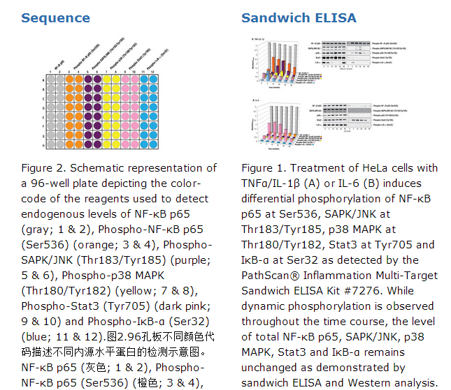
CST的PathScan® Inflammation Multi-Target Sandwich ELISA Kit #7276能够检测内源水平的六个蛋白: NF-κB p65, phospho-NF-κB p65 (Ser536), phospho-SAPK/JNK (Thr183/Tyr185), phospho-p38 MAPK (Thr180/Tyr182), phospho-Stat3 (Tyr705)和phospho-IκB-α (Ser32)。如图1所示,用多种细胞因子处理后能观察到随时间推移这些蛋白的不同激活方式。细胞的裂解液蛋白浓度与450 nm吸光度之间的关系在与单个的PathScan® Sandwich ELISA kits**相关的数据页中。**参考配套产品。此试剂盒能够检测已经经内部严格测试的物种的蛋白,但是也不排除检测到其它物种中同源性的蛋白。CST的PathScan® Inflammation Multi-Target Sandwich ELISA Kit是固相夹心酶联免疫吸收剂检测试剂盒(ELISA),试剂盒中包含检测内源水平NF-κB p65, phospho-NF-κB p65 (Ser536), phospho-SAPK/JNK (Thr183/Tyr185), phospho-p38 MAPK (Thr180/Tyr182), phospho-Stat3 (Tyr705) and phospho-IκB-α (Ser32)蛋白所需的试剂。这些分子代表了控制胁迫和炎症信号通路中重要的调节蛋白和结点蛋白。为每个目标蛋白提供了十六次测试。对已经标明的目标蛋白特异的实验验证能够在与单个PathScan® Sandwich ELISA Kits**相关的数据页中找到。简单的讲, 一个扑捉抗体*包裹到微孔的表面。与细胞裂解液孵育后, 微孔表面的抗体捕捉目标蛋白。经过严格的冲洗后,检测抗体*加入后检测已经捕捉到的目标蛋白。HRP连接的二抗加入用于识别检测抗体的结合。HRP的底物TMB加入进行显色反应。对反应的颜色,吸光度的数值按比例换算成目标蛋白的量。*试剂盒中的抗体是经过客户验证的特异性抗体。 **见配套产品。核转录因子κ B(NF-κB)/Rel 家族在炎症反应和免疫反应中发挥了至关重要的作用(1,2)。在哺乳动物中一共有5个家族 : RelA, c-Rel, RelB, NF-κB1 (p105/p50), 和 NF-κB2 (p100/p52)。这些蛋白以二聚体转录因子形式发挥作用。在未受到刺激的细胞中, NF-κB/Rel蛋白被隔离在细胞质中并被IκB蛋白所抑制 。NF-κB激活因子包括磷酸化的IκB蛋白,目的是降解它们并释放NF-κB/Rel复合体。激活的NF-κB/Rel 复合体通过磷酸化进一步的激活(1-4)。胁迫激活的蛋白激酶Jun N-末端激酶SAPK/JNK是通过环境中一系列的胁迫而激活,包括UV和gamma射线,神经酰胺,炎症因子、在某些情况下通过生长因子和GPCR激动剂(5-10)。与其它的MAPKs类似, 信号的中心单元是MAPKKK, 通常是MEKK1-4或者是混合的同谱系激酶(MLK), 它能够磷酸化并激活MKK4-7,然后磷酸化Thr183和Tyr185位点激活SAPK/JNK激酶(6)。胁迫信号是通过 Rho家族(Rac, Rho, cdc42)的小GTPases而传递到此级联反应(7)。Rac1和cdc42介导对MEKKs和MLKs的刺激(7)。或者作为另一种选择, MKK4-7 能够不依赖小GTPases而是通过刺激幼芽的中心激酶(GCK)家族中的一员而激活(8)。
p38 MAP 激酶(MAPK)参与到级联反应中控制细胞对促炎症因子和多种细胞胁迫的反应。MKK3,MKK6和SEK(MKK4)通过磷酸化Thr180和Tyr182位点激活p38 MAP激酶(11-14)。
Stat3转录因子是很多因子和生长因子受体的重要的信号分子。Stat3通过Tyr705位点的磷酸化而激活并引起二聚化,核定位和DNA结合(15,16)。
原厂资料:
Specificity / Sensitivity
CST's PathScan® Inflammation Multi-Target Sandwich ELISA Kit #7276 detects endogenous levels of six proteins: NF-κB p65, phospho-NF-κB p65 (Ser536), phospho-SAPK/JNK (Thr183/Tyr185), phospho-p38 MAPK (Thr180/Tyr182), phospho-Stat3 (Tyr705) and phospho-IκB-α (Ser32). Differential activation of these proteins can be observed over time in response to various cytokine treatments, as shown in Figure 1. The relationship between the protein concentration of the lysate and the absorbance at 450 nm can be found in the datasheets associated with the individual PathScan® Sandwich ELISA kits**. **See companion products. This kit detects proteins from the indicated species, as determined through in-house testing, but may also detect homologous proteins from other species.
Description
CST's PathScan® Inflammation Multi-Target Sandwich ELISA Kit is a solid phase sandwich enzyme-linked immunosorbent assay (ELISA) that combines the reagents necessary to detect endogenous levels of NF-κB p65, phospho-NF-κB p65 (Ser536), phospho-SAPK/JNK (Thr183/Tyr185), phospho-p38 MAPK (Thr180/Tyr182), phospho-Stat3 (Tyr705) and phospho-IκB-α (Ser32). These molecules represent convergence points and key regulatory proteins in signaling pathways controlling the stress and inflammation response. Sixteen tests are provided for each target protein. Specific assay formulations for the indicated target proteins can be found in the datasheets associated with the individual PathScan® Sandwich ELISA Kits**. Briefly, a capture antibody* has been coated onto the microwells. After incubation with cell lysates, the coated antibody captures the target protein. Following extensive washing, a detection antibody* is added to detect the captured target protein. An HRP-linked secondary antibody is then used to recognize the bound detection antibody. HRP substrate, TMB, is added to develop color. The magnitude of absorbance for this developed color is proportional to the quantity of bound target protein. *Antibodies in kit are custom formulations specific to kit. **See companion products.
Background
Transcription factors of the nuclear factor κB (NF-κB)/Rel family play a pivotal role in inflammation, stress and immune responses. There are five family members in mammals: RelA/p65, c-Rel, RelB, NF-κB1 (p105/p50) and NF-κB2 (p100/p52). These proteins function as dimeric transcription factors. In unstimulated cells, NF-κB/Rel proteins are sequestered in the cytoplasm and inhibited by the IκB proteins. NF-κB-activating agents induce phosphorylation of IκB's, targeting them for degradation and thereby releasing the NF-κB/Rel complexes. Active NF-κB/Rel complexes are further activated by phosphorylation (1-4).
The stress-activated protein kinase/Jun-amino-terminal kinase SAPK/JNK is activated by a variety of environmental stresses, including UV and gamma radiation, ceramides, inflammatory cytokines and in some instances, by growth factors and GPCR agonists (5-10). As with the other MAPKs, the core-signaling unit is composed of a MAPKKK, typically MEKK1-4, or by a mixed lineage kinase (MLK), which phosphorylates and activates MKK4-7, which then phosphorylates Thr183 and Tyr185 to activate the SAPK/JNK kinase (6). Stress signals are delivered to this cascade by small GTPases of the Rho family (Rac, Rho, cdc42) (7). Both Rac1 and cdc42 mediate the stimulation of MEKKs and MLKs (7). Alternatively, MKK4-7 can be activated by a pathway independent of small GTPases via stimulation of a member of the germinal center kinase (GCK) family (8).
p38 MAP kinase (MAPK) participates in a signaling cascade controlling the cellular response to pro-inflammatory cytokines and a variety of cellular stresses. MKK3, MKK6 and SEK (MKK4) activate p38 MAP kinase by phosphorylation at Thr180 and Tyr182 (11-14).
The Stat3 transcription factor is an important signaling molecule for many cytokines and growth factor receptors. Stat3 is activated by phosphorylation at Tyr705, which induces dimerization, nuclear translocation and DNA binding (15,16).
注意事项:
* 12 8-well modules -Each module is designed to break apart for 8 tests.
**Kit should be stored at 4°C with the exception of 10X Cell Lysis Buffer, which is stored at –20°C (packaged separately).


 京公网安备11010802025653 版权所有:北京逸优科技有限公司
