其它组分:
ELISA Sample Diluent【包装:25 ml,,运保温度:4°C】
STOP Solution 【子货号:#7002,包装:11 ml,,运保温度:4°C】
TMB Substrate 【子货号:#7004,包装:11 ml,,运保温度:4°C】
ELISA Wash Buffer (20X)【包装:25 ml,,运保温度:4°C】
Cell Lysis Buffer (10X) 【子货号:#9803,包装:15 ml,,运保温度:–20°C】
Sealing Tape【包装:2 sheets,,运保温度:4°C】
Anti-mouse IgG, HRP-linked Antibody【包装:1.8 ml,,运保温度:4°C】
Phospho-Akt (S473) Rabbit Antibody Coated Microwells【包装:16 tests,,运保温度:4°C】
Anti-mouse IgG, HRP-linked Antibody【包装:1.8 ml,,运保温度:4°C】
Akt1 Mouse Detection Antibody【包装:1.8 ml,,运保温度:4°C】
p38 MAPK Rabbit Detection Antibody【包装:1.8 ml,,运保温度:4°C】
Phospho-p38 MAPK (T180/Y182) Mouse Antibody Coated Microwells【包装:16 tests,,运保温度:4°C】
Anti-rabbit IgG, HRP-linked Antibody【包装:1.8 ml,,运保温度:4°C】
MEK1 Mouse Antibody Coated Microwells【包装:16 tests,,运保温度:4°C】
NF-κB p65 Rabbit Detection Antibody【包装:1.8 ml,,运保温度:4°C】
Phospho-NF-κB p65 (S536) Mouse Antibody Coated Microwells【包装:16 tests,,运保温度:4°C】
Anti-mouse IgG, HRP-linked Antibody【包装:1.8 ml,,运保温度:4°C】
Stat3 Rabbit Antibody Coated Microwells【包装:16 tests,,运保温度:4°C】
Akt Rabbit Antibody Coated Microwells【包装:16 tests,,运保温度:4°C】
Akt1 Mouse Detection Antibody【包装:1.8 ml,,运保温度:4°C】
Anti-rabbit IgG, HRP-linked Antibody【包装:1.8 ml,,运保温度:4°C】
Phospho-MEK1/2 (S217/221) Rabbit Detection Antibody【包装:1.8 ml,,运保温度:4°C】
Anti-rabbit IgG, HRP-linked Antibody【包装:1.8 ml,,运保温度:4°C】
Phospho-Stat3 (Y705) Mouse Detection Antibody【包装:1.8 ml,,运保温度:4°C】
描述:
CST的 PathScan® Signaling Nodes Multi-Target Sandwich ELISA Kit #7272能检测内源性六种蛋白水平:Akt1, phospho-Akt1 (Ser473), phospho-MEK1 (Ser217/221), phospho-p38 MAPK (Thr180/Tyr182), phospho-Stat3 (Tyr705) 和phospho-NF-κB p65 (Ser536)。如图1所示,经过一段时间对各种生长因子和细胞因子处理的应答能观察到这些蛋白的不同磷酸化。与每个PathScan® Sandwich ELISA Kits**有关的数据表中显示了裂解物蛋白浓度和450 nm处光吸收度之间的关系。**详见相关产品。该试剂盒经公司内部实验证明能检测已表明物种的蛋白,但也许能检测其他物种中的类似蛋白。CST的PathScan® Signaling Nodes Multi-Target Sandwich ELISA Kit是一种固相三明治酶联免疫吸附实验 (ELISA) ,它能测定内源性Akt1蛋白, 丝氨酸(473位点)磷酸化的Akt1, 丝氨酸(217/221)磷酸化的MEK1蛋白, 苏氨酸(180位点)/酪氨酸(182位点)磷酸化的p38 MAPK蛋白, 酪氨酸(705位点)磷酸化的Stat3蛋白和丝氨酸(536位点)磷酸化的NF-κB p65蛋白水平。这些分子代表了聚合点和信号转导通路中的关键调节蛋白,这些蛋白能控制细胞活动,如生长、分化、能量平衡和应答应激反应、炎性反应。对每个靶蛋白一共进行了16项测试。在有关个体的PathScan® Sandwich ELISA Kits**数据表中能找到已鉴定靶蛋白的特异性实验方法。总之,捕获抗体*能被覆盖在微量培养板上。用细胞裂解液孵化后,靶蛋白就能被原来覆盖的抗体捕获。接着反复清洗后加入足量的p44/42 MAPK mouse detection mAb以检测捕获的phospho-p44/42 MAPK蛋白。然后用HRP-linked二抗识别已绑定的检测性抗体。加入HRP的底物——TMB用于显色。显色剂颜色的深浅与限制的靶蛋白的含量成比例。这个试剂盒中的*抗体是特异定制的。Akt是一种原癌基因,它对各种细胞进程起着重要的调节作用,包括生长、生存和细胞周期。 Akt蛋白也是胰岛素信号转导和葡萄糖代谢的重要调节子(1-4)。Akt蛋白被PI3激酶信号转导活化。活化环内的苏氨酸(308位点)被PDK1磷酸化,蛋白羧端丝氨酸(473位点)被PDK2磷酸化,这两个位点的磷酸化也使Akt蛋白被激活(5-7)。MEK1、 MEK2是一种双特异性蛋白激酶,在有丝分裂原活化的蛋白激酶级联中控制细胞生长和分化。MEK1、 MEK2的活化通过两个丝氨酸(217、221位点)被Raf样分子磷酸化而发生。MEK蛋白能活化p44 and p42 MAP激酶(8-10)。p38 MAP kinase (MAPK)参与信号转导的级联反应,控制细胞应答前炎性细胞因子各种细胞应激。MKK3, MKK6,SEK (MKK4)通过苏氨酸(180位点)和酪氨酸(182位点)的磷酸化而激活 p38 MAP激酶(11-14)。Stat3蛋白转录因子是许多细胞因子生长因子受体的重要信号转导分子。Stat3蛋白酪氨酸(705位点)磷酸化后就被激活,诱导二聚化、胞核内转位和DNA结合(15,16)。细胞核因子κB (NF-κB)/Rel家族的转录因子在炎性反应、应激反应和免疫应答中扮演着重要角色。哺乳动物中的五类家族成员:RelA/p65, c-Rel, RelB, NF-κB1 (p105/p50) and NF-κB2 (p100/p52)。这些蛋白作为二聚体转录因子发挥作用。在非刺激细胞中NF-κB/Rel蛋白隐蔽在胞质中,并被IκB蛋白抑制。NF-κB-活化试剂能诱导IκB的磷酸化,将它们作为降解的靶点,因此能释放NF-κB/Re复合物。活化的NF-κB/Re复合物被磷酸化后会进一步活化(17-20)。
原厂资料:
Specificity / Sensitivity
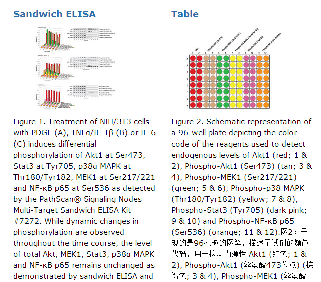
CST's PathScan® Signaling Nodes Multi-Target Sandwich ELISA Kit #7272 detects endogenous levels of six proteins: Akt1, phospho-Akt1 (Ser473), phospho-MEK1 (Ser217/221), phospho-p38 MAPK (Thr180/Tyr182), phospho-Stat3 (Tyr705) and phospho-NF-κB p65 (Ser536). Differential phosphorylation of these proteins can be observed over time in response to various growth factor and cytokine treatments, as shown in Figure 1.
The relationship between the protein concentration of the lysate and the absorbance at 450 nm can be found in the datasheets associated with the individual PathScan® Sandwich ELISA Kits**. **See companion products.
This kit detects proteins from the indicated species, as determined through in-house testing, but may also detect homologous proteins from other species.
Description
CST's PathScan® Signaling Nodes Multi-Target Sandwich ELISA Kit is a solid phase sandwich enzyme-linked immunosorbent assay (ELISA) that combines the reagents necessary to detect endogenous levels of Akt1, phospho-Akt1 (Ser473), phospho-MEK1 (Ser217/221), phospho-p38 MAPK (Thr180/Tyr182), phospho-Stat3 (Tyr705) and phospho-NF-κB p65 (Ser536). These molecules represent convergence points and key regulatory proteins in signaling pathways controlling cellular events such as growth and differentiation, energy homeostasis, and the response to stress and inflammation. Sixteen tests are provided for each target protein. Specific assay formulations for the indicated target proteins can be found in the datasheets associated with the individual PathScan® Sandwich ELISA Kits**. Briefly, a capture antibody* has been coated onto the microwells. After incubation with cell lysates, the target protein is captured by the coated antibody. Following extensive washing, a detection antibody* is added to detect the captured target protein. An HRP-linked secondary antibody is then used to recognize the bound detection antibody. HRP substrate, TMB, is added to develop color. The magnitude of absorbance for this developed color is proportional to the quantity of bound target protein. *Antibodies in kit are custom formulations specific to kit. **See companion products.
Background
Akt is a protooncogene with a critical regulatory role in diverse cellular processes including growth, survival and the cell cycle. Akt is also a major regulator of insulin signaling and glucose metabolism (1-4). Akt is activated by PI3 kinase signaling and activation loop phosphorylation at Thr308 by PDK1 and by phosphorylation withing the carboxyl terminus at Ser473 by PDK2 (5-7).
MEK1 and MEK2 are dual-specificity protein kinases that function in a mitogen activated protein kinase cascade controlling cell growth and differentiation. Activation of MEK1 and MEK2 occurs through phosphorylation of serine 217 and serine 221 by Raf-like molecules. MEK activates p44 and p42 MAP kinase (8-10).
p38 MAP kinase (MAPK) participates in a signaling cascade controlling the cellular response to pro-inflammatory cytokines and a variety of cellular stresses. MKK3, MKK6 and SEK (MKK4) activate p38 MAP kinase by phosphorylation at Thr180 and Tyr182 (11-14).
The Stat3 transcription factor is an important signaling molecule for many cytokines and growth factor receptors. Stat3 is activated by phosphorylation at Tyr705, which induces dimerization, nuclear translocation and DNA binding (15,16).
Transcription factors of the nuclear factor κB (NF-κB)/Rel family play a pivotal role in inflammation, stress and immune responses. There are five family members in mammals: RelA/p65, c-Rel, RelB, NF-κB1 (p105/p50) and NF-κB2 (p100/p52). These proteins function as dimeric transcription factors. In unstimulated cells, NF-κB/Rel proteins are sequestered in the cytoplasm and inhibited by the IκB proteins. NF-κB-activating agents induce phosphorylation of IκB's, targeting them for degradation and thereby releasing the NF-κB/Rel complexes. Active NF-κB/Rel complexes are further activated by phosphorylation (17-20).


 京公网安备11010802025653 版权所有:北京逸优科技有限公司
