Phospho-FGF Receptor (Tyr653/654) (55H2) Mouse mAb detects transfected levels of FGF receptors only when phosphorylated at tyrosines 653/654. The antibody cross-reacts slightly with activated PDGF and insulin/IGF-I receptors.
Source / Purification
Monoclonal antibody is produced by immunizing animals with a synthetic phosphopeptide corresponding to residues surrounding Tyr653/654 of human FGF receptor-1. The corresponding sequence is identical in FGF receptor-2, -3 and -4.
Background
Fibroblast growth factors (FGFs) produce mitogenic and angiogenic effects in target cells by signaling through cell surface receptor tyrosine kinases. There are four members of the FGF receptor family: FGFR-1 (flg), FGFR-2 (bek, KGFR), FGFR-3, and FGFR-4. Each receptor contains an extracellular ligand binding domain, a transmembrane domain, and a cytoplasmic kinase domain (1). Following ligand binding and dimerization, the receptors are phosphorylated at specific tyrosine residues (2). Seven tyrosine residues in the cytoplasmic tail of FGFR-1 can be phosphorylated: Tyr463, 583, 585, 653, 654, 730, and 766. Tyr653 and Tyr654 are important for catalytic activity of activated FGFR and are essential for signaling (3). The other phosphorylated tyrosine residues may provide docking sites for downstream signaling components such as Crk and PLCγ (4,5).
Storage:Supplied in 10 mM sodium HEPES (pH 7.5), 150 mM NaCl, 100 µg/ml BSA, 50% glycerol and less than 0.02% sodium azide. Store at –20°C. Do not aliquot the antibody.
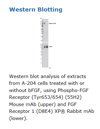